AskAlth is an independent educational resource created by NHS doctors and critical care nurses. It is not affiliated with CC3N, NICE, or the NHS, though our content is mapped to and referenced against their published frameworks and guidance.

Heart Valves
How to use this resource, and learning outcomes:
1. Why the heart valves matter at the bedside
2. Overview: the four valves
3. The atrioventricular valves
4. The semilunar valves
5. The fibrous skeleton and leaflet structure
6. Valves are passive: pressure gradients open and close them
7. The aortic root and coronary perfusion
8. Hearing the valves: heart sounds and auscultation
9. Valve movement and the waveforms you monitor
10. Valves and positive pressure ventilation
11. Clinical implications for the acute care nurse
Abbreviations
12. Self-test
Glossary
Recommended pages
Further reading and local governance
What this page covers — and what it does not
This page covers the NORMAL heart valve: where each valve sits, what it is made of, how it opens and closes, where to listen to it, and how valve movement produces the waveforms on your monitor.
For valve DISEASE — stenosis and regurgitation, causes, murmurs of each lesion, severity grading, haemodynamic targets, endocarditis, prosthetic valves, anticoagulation and post-operative care — see Acquired Heart Valve Disease.
Work through this page first. The disease page assumes the anatomy and physiology below.
How to use this resource
Sections 2 to 7 build the anatomy and physiology. Sections 8 to 11 apply it: what you hear through a stethoscope, what valve movement is doing to the arterial and CVP traces in front of you, and how positive pressure ventilation changes valve loading.
ICU Competencies:
If you are working through Step 1, sections 2 to 8 and 11 are your priority.
If you are working through Step 2 or an academic critical care programme, sections 9 and 10 are the rationale sections on this page — and Acquired Heart Valve Disease carries the rest.
Learning outcomes:
Step 1 — by the end of this session you will be able to:
Name the four cardiac valves, state their position and describe their structure and supporting apparatus.
Explain that valves are passive structures driven by pressure gradients, and relate each valve’s opening and closing to the cardiac cycle.
Describe the atrial kick and explain why atrial fibrillation is poorly tolerated in a non-compliant ventricle.
Describe the function of the fibrous skeleton, including its role in electrical insulation.
Locate the four auscultation areas and state which valves close to produce S1 and S2.
Explain why the left coronary tree fills during diastole and what determines coronary perfusion pressure.
Step 2 — in addition, you will be able to:
Identify the point of aortic valve closure on an arterial waveform and explain what it represents.
Relate the a, c and v waves of the CVP trace to the movement of the tricuspid valve.
Explain the effects of positive pressure ventilation on preload and afterload, and predict which valve loading states tolerate it badly.
Explain, using coronary perfusion pressure, why tachycardia threatens the hypertrophied ventricle.
1. Why the heart valves matter at the bedside
The heart valves ensure one-way blood flow through the chambers. They are passive structures — they open and close in response to the pressure gradient across them during the cardiac cycle, not because of any muscular action of their own.
Valves explain the numbers on your monitor. The dicrotic notch is aortic valve closure. The v wave on a CVP trace is the atrium filling against a shut tricuspid valve. If you know what the valves are doing, the waveform stops being decoration and starts being information.
Valves explain what you hear. Every heart sound is a valve closing, and every murmur is turbulent flow through a valve that is not doing its job. The bedside nurse is usually the first person placed to notice that a murmur was not there yesterday.
Valve anatomy explains valve emergencies. The blood supply to one papillary muscle, and the avascular structure of the leaflets, between them account for two of the ways a valve fails suddenly. Both are covered here anatomically and on Acquired Heart Valve Disease clinically.
2. Overview: the four valves
There are four valves: two atrioventricular (AV) valves separating atria from ventricles, and two semilunar valves separating ventricles from the great arteries.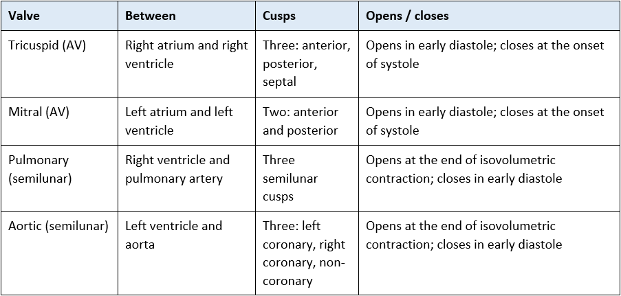

3. The atrioventricular valves
3.1 Tricuspid valve
Location: between the right atrium and right ventricle.
Structure: three cusps — anterior, posterior and septal.
Function: opens in early diastole when falling right ventricular pressure drops below right atrial pressure, allowing the ventricle to fill. Closes at the onset of systole when ventricular pressure exceeds atrial pressure, preventing backflow.
Support: chordae tendineae attach the free edges of the cusps to three papillary muscles, which contract at the start of systole to tension the chordae and stop the cusps prolapsing into the atrium.
3.2 Mitral valve
Location: between the left atrium and left ventricle.
Structure: two leaflets, anterior and posterior. The posterior leaflet is conventionally divided into three scallops (P1, P2, P3) with corresponding anterior segments (A1, A2, A3). You will see this terminology in echocardiography reports.
Support: chordae tendineae and two papillary muscles — anterolateral and posteromedial.
A note on terminology
The mitral valve is sometimes called the “bicuspid valve”. This is anatomically accurate but best avoided in clinical writing, because bicuspid aortic valve is a distinct congenital variant — present in roughly 1 to 2% of the population and a leading cause of aortic stenosis in adults under 65.
3.3 The atrial kick
Ventricular filling is largely passive. Atrial contraction occurs late in diastole and contributes only the final 20 to 30% of ventricular filling — the atrial kick.
When a patient goes into atrial fibrillation this contribution is lost, along with the regularity of filling. In a healthy compliant ventricle the effect may be modest. In a stiff, hypertrophied or poorly compliant ventricle — aortic stenosis, mitral stenosis, hypertrophic cardiomyopathy, diastolic dysfunction — the ventricle depends heavily on that final filling, and the loss can precipitate abrupt hypotension and pulmonary oedema.
Practice point: this is why restoring or controlling rhythm is a haemodynamic intervention in these patients, not just a rhythm-strip tidy-up, and why new atrial fibrillation in a patient with known valve disease warrants prompt escalation.
3.4 Papillary muscles and their blood supply
The anterolateral papillary muscle usually has a dual blood supply, from the left anterior descending and circumflex arteries.
The posteromedial papillary muscle typically has a single supply, from the posterior descending artery.
That single blood supply makes the posteromedial papillary muscle vulnerable in inferior myocardial infarction. Rupture or dysfunction causes acute severe mitral regurgitation — a surgical emergency. For the presentation, why the murmur may be soft or absent, and what to do, see Acquired Heart Valve Disease.
4. The semilunar valves
4.1 Pulmonary valve
Between the right ventricle and the pulmonary artery; three crescent-shaped semilunar cusps. It opens when right ventricular pressure exceeds pulmonary artery diastolic pressure — typically around 8 to 12 mmHg — at the end of isovolumetric contraction, and closes when the gradient reverses in early diastole.
4.2 Aortic valve
Between the left ventricle and the aorta; three semilunar cusps named left coronary, right coronary and non-coronary. It opens when left ventricular pressure exceeds aortic diastolic pressure — typically around 80 mmHg — and closes when aortic pressure exceeds falling ventricular pressure.
Because the pulmonary valve opens slightly earlier and closes slightly later than the aortic valve, the second heart sound shows physiological splitting, which widens on inspiration as increased venous return prolongs right ventricular ejection.
5. The fibrous skeleton and leaflet structure
5.1 The fibrous skeleton (annuli fibrosi)
All four valves sit within a ring of dense connective tissue at the base of the ventricles. It has three functions:
Anchoring — provides the annulus for each valve and an attachment point for atrial and ventricular myocardium.
Electrical insulation — separates atrial from ventricular myocardium, so the AV node and bundle of His form the only normal conducting pathway between them. See Cardiac Conduction System.
Maintaining annular geometry — keeping the leaflets in a position where they can coapt, that is meet and seal.
When a chamber dilates, the annulus stretches and the leaflets no longer coapt properly. This produces functional (secondary) regurgitation with structurally normal leaflets — the mechanism behind functional mitral regurgitation in dilated cardiomyopathy and functional tricuspid regurgitation in pulmonary hypertension.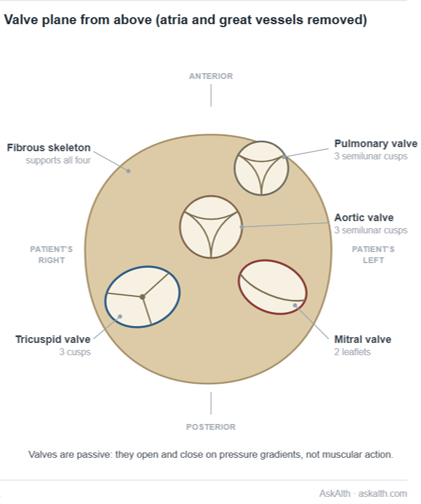
5.2 Leaflet structure
Each leaflet is a three-layered, largely avascular structure covered by endothelium. Because leaflets have no significant blood supply of their own and are bathed directly in blood, they have limited immune surveillance and poor antibiotic penetration — the anatomical reason infective endocarditis is difficult to treat and may require surgery. See Acquired Heart Valve Disease.
The three layers are: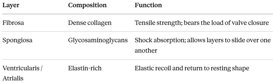
6. Valves are passive: pressure gradients open and close them
No valve has muscle of its own. Each opens when pressure upstream exceeds pressure downstream, and closes when that gradient reverses. Everything else — heart sounds, waveforms, murmurs — follows from that.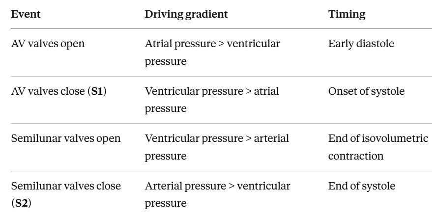
The sequence in one beat
Late diastole: AV valves open, semilunar valves shut. Ventricles fill, then the atria contract (the atrial kick).
Onset of systole: ventricular pressure rises above atrial pressure. AV valves shut — this is S1. All four valves are now closed: isovolumetric contraction.
Ventricular pressure exceeds arterial pressure: semilunar valves open. Ejection.
End of systole: ventricular pressure falls below arterial pressure. Semilunar valves shut — this is S2. All four valves closed again: isovolumetric relaxation.
Ventricular pressure falls below atrial pressure: AV valves open and filling begins again.
See The Cardiac Cycle.
7. The aortic root and coronary perfusion
The aortic valve sits within the aortic root. Its three outward bulges — the sinuses of Valsalva — contain the left and right coronary ostia. Eddy currents within the sinuses help the cusps close cleanly and keep the ostia from being obstructed by the open cusps.
Because the left ventricle compresses its own myocardium during systole, the left coronary tree fills during diastole, driven by aortic root pressure after the aortic valve has closed.
Coronary perfusion pressure ≈ aortic diastolic pressure − left ventricular end-diastolic pressure (LVEDP)
Two practical consequences:
A hypertrophied ventricle can be ischaemic with normal coronary arteries. LVEDP is raised and muscle mass is increased, so supply falls short of demand even with unobstructed vessels.
Tachycardia shortens diastole disproportionately, reducing both coronary filling time and ventricular filling time. This is why tachyarrhythmias are so badly tolerated where the ventricle is stiff or the valve is narrowed.
See Coronary Artery Anatomy for the wider coronary circulation.
8. Hearing the valves: heart sounds and auscultation
Auscultation areas are the points on the chest wall where sound from each valve is best transmitted. They do not sit directly over the anatomical valve — they lie downstream, in the direction of blood flow.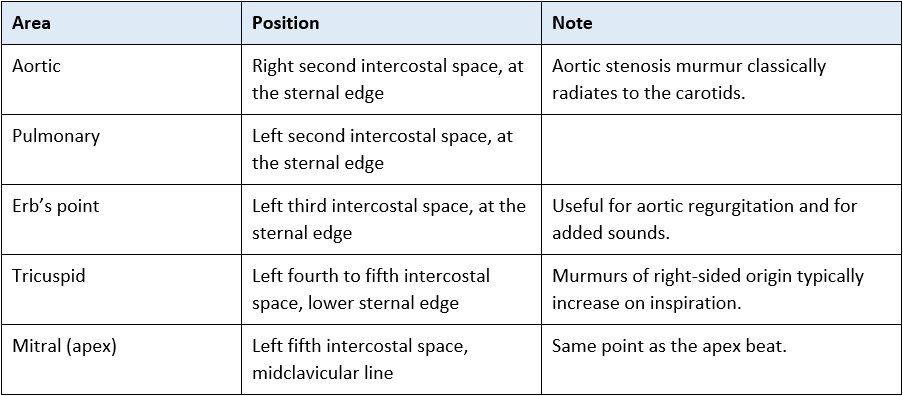
The heart sounds
S1 — closure of the mitral and tricuspid valves at the onset of systole.
S2 — closure of the aortic and pulmonary valves at the onset of diastole, with physiological splitting that widens on inspiration.
S3 — early diastolic, caused by rapid ventricular filling into a volume-loaded or poorly compliant ventricle. Normal in the young and in pregnancy; in an older critically ill patient it suggests fluid overload or heart failure.
S4 — late diastolic, caused by atrial contraction against a stiff ventricle. It cannot occur in atrial fibrillation, because there is no organised atrial contraction.
Practice point — auscultation in a noisy critical care unit
Ventilator noise, filtration circuits and alarms make subtle murmurs very difficult to hear. Reduce what you can, position the patient where safe, and use the diaphragm for high-pitched sounds and the bell lightly applied for low-pitched ones.
Document what you heard, where, and when — and compare it with the previous shift.
For the characteristic murmur of each valve lesion, see Acquired Heart Valve Disease. For technique in detail, see Heart Sounds (auscultation).
9. Valve movement and the waveforms you monitor
Step 2 content — rationale and application
Both the arterial and central venous traces are direct recordings of valve movement and its consequences. Reading them this way makes them far easier to interpret.
9.1 The arterial waveform
The upstroke begins when the aortic valve opens and ejection starts.
The dicrotic notch is aortic valve closure — the brief backflow towards the ventricle that snaps the cusps shut. It marks the end of systole on the trace.
The diastolic decay that follows is the runoff of blood into the systemic circulation, and it is the pressure that drives coronary filling — see section 7.
A slurred, damped or absent dicrotic notch is worth noticing. Check the transducer, tubing, flush system and any air bubbles first, because over-damping produces the same appearance. If the system is sound, report it.
9.2 The CVP waveform
The a, c and v waves are the tricuspid valve and the right atrium telling you what they are doing.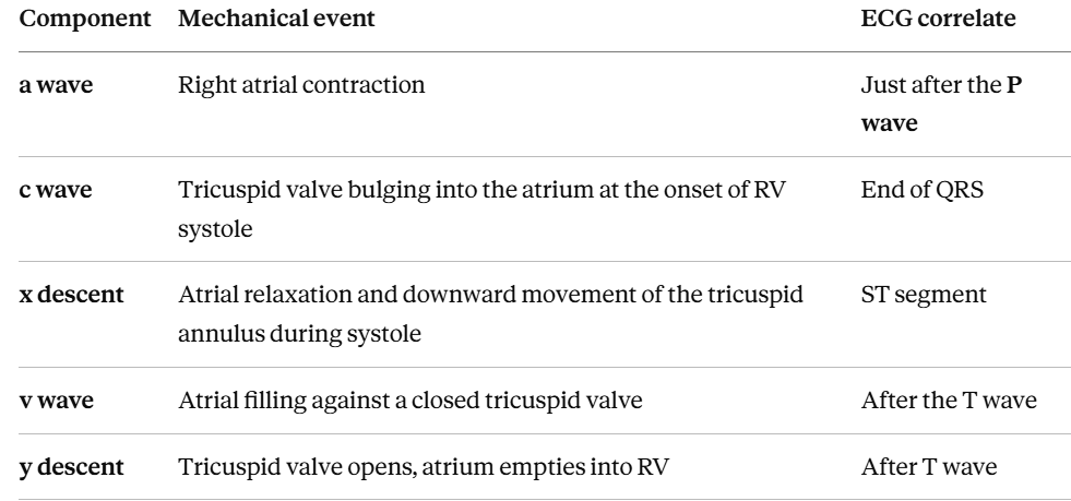
Valve lesions distort these waveforms in characteristic ways, and there are situations in which cardiac output measurement becomes unreliable as a result. Those lesion-specific caveats are covered on Acquired Heart Valve Disease and on Invasive Monitoring – Waveforms.
10. Valves and positive pressure ventilation
Step 2 content — rationale and application
– Positive pressure ventilation raises intrathoracic pressure throughout the respiratory cycle, changing the loading conditions on both sides of every valve.
– Preload falls. Raised intrathoracic pressure reduces the pressure gradient for venous return to the right atrium, so less blood crosses the tricuspid valve.
– Left ventricular afterload falls. Raised intrathoracic pressure reduces transmural left ventricular pressure, so the ventricle ejects against less effective load across the aortic valve.
– Pulmonary vascular resistance changes with lung volume. It is lowest around functional residual capacity and rises with both atelectasis and overdistension, so high PEEP and high plateau pressures increase the load the right ventricle must overcome to open the pulmonary valve.
What to anticipate
Where filling is already marginal or obstructed, the fall in preload at induction and the switch to positive pressure is poorly tolerated and can be profound. Anticipate it, have vasopressor drawn up, and expect the team to induce cautiously.
Where the problem is pulmonary oedema from a failing left ventricle, positive pressure usually helps — non-invasive ventilation or CPAP reduces both preload and afterload and often produces rapid improvement.
Weaning reverses all of it. The return to negative pressure breathing increases venous return and left ventricular afterload and increases the work of breathing, which can precipitate weaning-induced pulmonary oedema.
For which specific lesions tolerate this badly and why, see Acquired Heart Valve Disease. See also Starling’s Law and Ventilator Weaning and Extubation – Overview.
11. Clinical implications for the acute care nurse
Assessment
Auscultate at all four areas as part of your cardiovascular assessment, and document what you heard and where.
Treat a new murmur, or a murmur that has changed in loudness or timing, as a finding to escalate rather than to record and move on from.
Note whether S3 or S4 is present — both carry information about ventricular filling and compliance.
Locate the apex beat where you can; it is both the mitral auscultation point and a marker of ventricular size and mediastinal position.
Monitoring
Look for the dicrotic notch on every arterial trace. Its position and clarity tell you about ejection and about the fidelity of your monitoring system.
Read the CVP waveform, not only the number. Absent a waves, prominent v waves and blunted descents all mean something.
Level and zero transducers properly — an incorrectly levelled transducer will mislead you about every one of the above.
Anticipating trouble
Protect rhythm in any patient with a stiff or hypertrophied ventricle. The atrial kick is worth 20 to 30% of filling and its loss can be abrupt and severe.
Treat tachycardia as a filling and coronary perfusion problem, not only a rhythm problem.
Anticipate a fall in blood pressure at induction and intubation, and after any increase in PEEP.
Escalate immediately for sudden deterioration with a new murmur, flash pulmonary oedema with shock, or a new murmur after myocardial infarction. See Acquired Heart Valve Disease for the specific emergencies and their management.
12. Self-test
Check your understanding. The section to check your answer against is shown in brackets.
Step 1
Name the four cardiac valves, state where each sits and how many cusps each has. (Section 2)
Why are the heart valves described as passive structures? (Sections 1 and 6)
What is the atrial kick, roughly how much does it contribute, and why does its loss matter more in a stiff ventricle? (Section 3.3)
Give the three functions of the fibrous skeleton. Why does the electrical insulation matter? (Section 5.1)
Which valves close to produce S1, and which produce S2? Where would you listen for the mitral valve? (Section 8)
Describe the sequence of valve opening and closing through one cardiac cycle. (Section 6)
Why does the left coronary tree fill during diastole rather than systole? (Section 7)
Which papillary muscle has a single blood supply, and which artery supplies it? (Section 3.4)
Step 2
Point to the dicrotic notch on an arterial trace and explain what it represents. What are the two commonest reasons it looks slurred? (Section 9.1)
Match the a, c and v waves of the CVP trace to what the tricuspid valve is doing. Why is the a wave absent in atrial fibrillation? (Section 9.2)
Explain, using coronary perfusion pressure, why a patient with a hypertrophied ventricle can be ischaemic with normal coronary arteries. (Section 7)
Your patient is about to be induced and ventilated. Predict what will happen to preload and left ventricular afterload, and explain the mechanism. (Section 10)
Why does raising PEEP sometimes worsen right ventricular performance? (Section 10)
Why can a patient move from sinus rhythm to atrial fibrillation with no change in rate and still become hypotensive? (Section 3.3)
Abbreviations
AF – Atrial fibrillation
AV – Atrioventricular
CC3N – Critical Care Networks – National Nurse Leads
CPAP – Continuous positive airway pressure
CVP – Central venous pressure
LVEDP – Left ventricular end-diastolic pressure
PEEP – Positive end-expiratory pressure
PVR – Pulmonary vascular resistance
S1–S4 – First to fourth heart sounds
Glossary
Annulus — the fibrous ring supporting a valve.
Coaptation — the meeting and sealing of valve leaflets when closed.
Cusp / leaflet — an individual flap of the valve.
Dicrotic notch — the deflection on the arterial waveform marking aortic valve closure.
Isovolumetric contraction / relaxation — the phases when all four valves are closed and ventricular volume does not change.
Prolapse — displacement of a leaflet back into the upstream chamber.
Sinuses of Valsalva — the three outward bulges of the aortic root containing the coronary ostia.
Recommended pages
Next in this section
The Cardiac Cycle — where valve movement fits in time
Heart Sounds (auscultation) — S1, S2, splitting and murmurs in detail
Structure and Function of the Heart
Cardiac Surface Anatomy — where to place your stethoscope
Cardiac Conduction System — the insulating role of the fibrous skeleton
When a valve fails
Acquired Heart Valve Disease — the companion page. Causes, the eight lesions, murmurs of each, severity, haemodynamic targets, endocarditis, prosthetic valves and post-operative care
Applying this clinically
Invasive Monitoring – Waveforms – Art, CVP, PA, PCWP; Arterial Line Transducer
Cardiac Output Monitoring; Starling’s Law
Coronary Artery Anatomy; Pathophysiology of Heart Failure
Basic Modes of Ventilation; Ventilator Weaning and Extubation – Overview
Further reading and local governance
Guidelines and reference
British Society of Echocardiography — minimum datasets and valve assessment protocols.
CC3N National Competency Framework for Registered Nurses in Adult Critical Care — Step 1 and Step 2.
2025 ESC/EACTS Guidelines for the Management of Valvular Heart Disease — for the disease context; see the companion page.
Local governance
Your local guidance on invasive haemodynamic monitoring, transducer levelling and zeroing.
Your local cardiovascular assessment and documentation standards.
Textbooks
Levick, An Introduction to Cardiovascular Physiology
Adam, Osborne and Welch, Critical Care Nursing: Science and Practice (Oxford)
Bersten and Handy, Oh’s Intensive Care Manual
Last reviewed: August 2026. Next review due: August 2027. AskAlth Nursing Team, London UK.
AskAlth is an educational resource for registered healthcare professionals in UK adult critical care. Not for patients or the public. Not a substitute for local policy, clinical guidance or the BNF. Content reflects guidance at the time of writing. AskAlth is independent and not affiliated with CC3N, NICE, the Resuscitation Council UK, the NMC or the NHS.
AskAlth is an independent educational resource created by NHS doctors and critical care nurses. It is not affiliated with CC3N, NICE, or the NHS, though our content is mapped to and referenced against their published frameworks and guidance.