

Cardiac Conduction System
How to use this resource, and learning outcomes:
1. Why the conduction system matters at the bedside
2. The sequence of conduction
3. The components
4. Automaticity and the pacemaker hierarchy
5. Autonomic control of rate and conduction
6. Blood supply to the conduction system
7. Linking the conduction system to the ECG
8. When conduction fails: blocks and escape rhythms
9. Reversible causes: electrolytes, drugs and stimuli
10. Electrical activity is not mechanical output
11. Pacing: transcutaneous, temporary and permanent
12. Clinical implications for the acute care nurse
Abbreviations
13. Self-test
Glossary
Recommended pages
Further reading and local governance
How to use this resource
Sections 2 to 6 build the anatomy and physiology. Sections 7 to 12 apply it: what each part of the conduction system contributes to the ECG in front of you, what happens when it fails, what commonly causes that failure, and what you do about it.
ICU Step Competencies:
If you are working through Step 1, sections 2 to 8, 9 and 12 are your priority.
If you are working through Step 2 or an academic critical care programme, sections 8 to 11 are where the rationale sits — you will be expected to explain why a block is dangerous, not simply name it.
Learning outcomes
Step 1 — by the end of this session you will be able to:
– Describe the normal cardiac conduction pathway in sequence, naming each component and its location.
– State the intrinsic rate of the sinoatrial node, the atrioventricular junction and the ventricular escape tissue, and explain the pacemaker hierarchy.
– Explain which part of the ECG complex corresponds to which part of the conduction pathway, including normal PR and QRS durations.
– Recognise first-degree, Mobitz I, Mobitz II and complete atrioventricular block on a rhythm strip and state which are dangerous.
– List the common reversible causes of bradycardia and conduction disturbance in critical care.
– Explain why a rhythm on the monitor must always be correlated with a pulse or arterial trace.
Step 2 — in addition, you will be able to:
– Give a rationale for why block at the atrioventricular node behaves differently from block below it, and why this changes the response to atropine.
– Relate the site of myocardial infarction to the type of conduction disturbance you should anticipate.
– Explain the haemodynamic rationale for preserving atrioventricular synchrony when pacing, and relate it to ventricular compliance.
– Interpret cannon a waves on the CVP or JVP and explain their mechanism.
Identify the electrolyte and pharmacological contributors to conduction disturbance in your own patients and evaluate the effect of correcting them.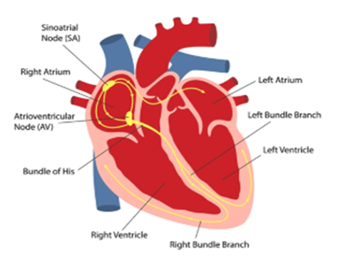
1. Why the conduction system matters at the bedside
The cardiac conduction system is the network of specialised muscle cells that generate and transmit electrical impulses, allowing the heart to contract in a coordinated, rhythmic sequence. It is what turns a bag of muscle into a pump.
Everything on your monitor is this system. The rhythm strip is not a picture of the heart beating — it is a picture of electrical conduction. Learning which part of the pathway produces which part of the complex is what turns rhythm recognition from memorising shapes into reasoning.
Conduction disturbance in critical care is usually caused by something you can fix. Hypoxia, potassium, magnesium, drugs, vagal stimuli, acidosis and ischaemia are all common and all correctable. The nurse at the bedside is usually the first to see the change and often the person who identifies the cause.
Some blocks are benign and some are pre-arrest. The difference between Mobitz I and Mobitz II is not a piece of exam trivia — it determines whether you continue observing or get the pacing equipment to the bedside.
2. The sequence of conduction
The sinoatrial (SA) node depolarises spontaneously.
The impulse spreads across both atria, producing atrial depolarisation and then atrial contraction.
The impulse reaches the atrioventricular (AV) node, where it is deliberately delayed.
It passes into the bundle of His, the only normal electrical connection between atria and ventricles.
It divides into the right and left bundle branches, the left dividing further into anterior and posterior fascicles.
It spreads through the Purkinje network, depolarising the ventricular myocardium rapidly and almost simultaneously, producing ventricular contraction.
Why the AV nodal delay exists
The delay is roughly 0.1 seconds, and it is deliberate. It allows atrial contraction to complete and top up ventricular filling — the atrial kick — before the ventricles contract.
The AV node also acts as a gatekeeper. Its slow recovery limits how many impulses can reach the ventricles, which is what protects the patient in atrial fibrillation and atrial flutter: the atria may be firing at 300 to 600 per minute, but only a fraction get through.
3. The components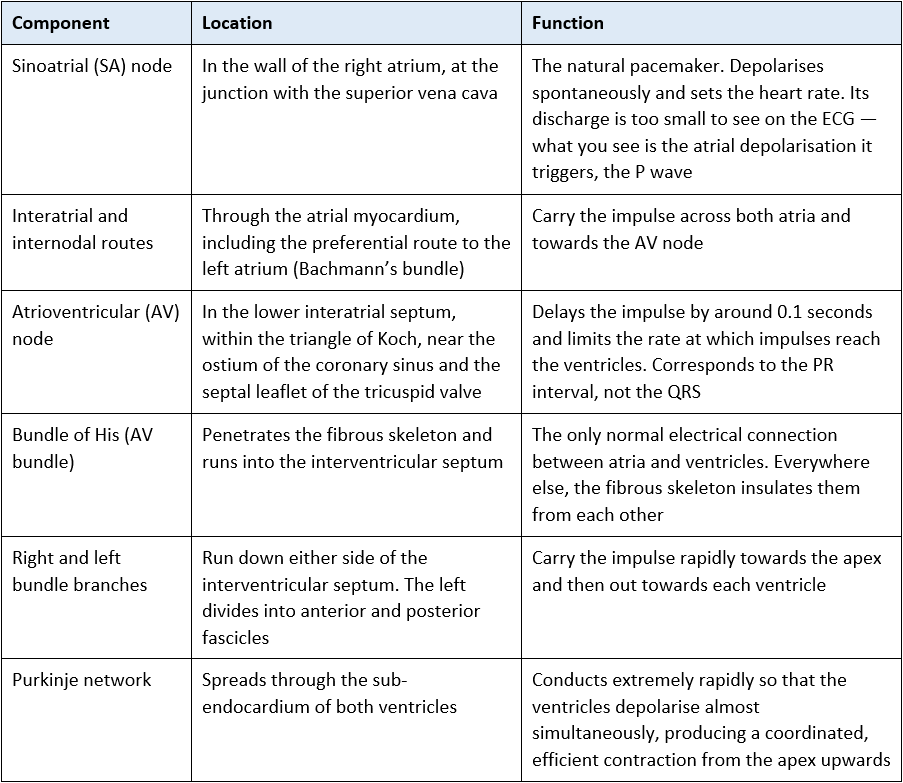
Cross-link: the insulating role of the fibrous skeleton is described on the Heart Valves page. It is the reason the bundle of His matters so much — if it or the branches below it fail, there is no alternative route.
4. Automaticity and the pacemaker hierarchy
Several tissues in the heart can depolarise spontaneously. The fastest one wins and suppresses the others — normally the SA node. If it fails, or if conduction from it is blocked, the next tissue down takes over at its own slower intrinsic rate. This is the escape hierarchy.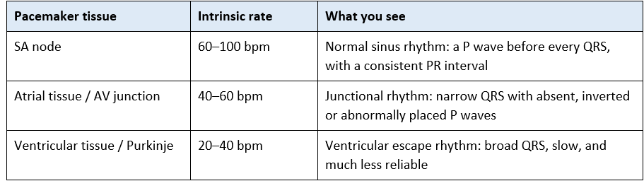
The clinical rule of thumb: the lower down the escape focus, the slower and less dependable the rhythm. A narrow-complex junctional escape at 50 is a patient you watch closely. A broad-complex ventricular escape at 25 is a patient who needs pacing equipment at the bedside now.
5. Autonomic control of rate and conduction
Parasympathetic (vagal) supply acts mainly on the SA and AV nodes through muscarinic (M2) receptors. It slows the rate of SA nodal firing and slows AV nodal conduction. Vagal tone is dominant at rest, which is why the resting rate is below the SA node’s intrinsic rate.
Sympathetic supply acts through beta-1 receptors across the whole conduction system and myocardium, increasing rate, speeding AV conduction and increasing contractility.
Vagal stimuli you will create yourself
Tracheal suctioning, laryngoscopy and intubation, nasogastric tube insertion, straining or vomiting, abdominal insufflation, pressure on the eye, and raised intracranial pressure (Cushing’s reflex) can all provoke profound bradycardia.
Anticipate it: monitor throughout the procedure, keep the pause short, pre-oxygenate before suctioning, and stop and re-oxygenate if the rate falls.
Have atropine and the emergency trolley available for high-risk patients, and know your local policy. See Management of Airway Secretions and Endotracheal Intubation.
6. Blood supply to the conduction system
The SA node is supplied by the SA nodal artery — from the right coronary artery in roughly 60% of people and the left circumflex in roughly 40%.
The AV node is supplied by the AV nodal artery, which arises from whichever vessel gives the posterior descending artery — the right coronary artery in the majority (right-dominant) pattern.
The bundle branches are largely supplied by septal perforators from the left anterior descending artery.
Infarct territory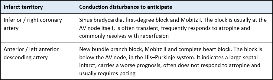
See Coronary Artery Anatomy for the wider coronary circulation and dominance.
7. Linking the conduction system to the ECG
At the standard paper speed of 25 mm per second, each small square is 0.04 seconds and each large square is 0.2 seconds.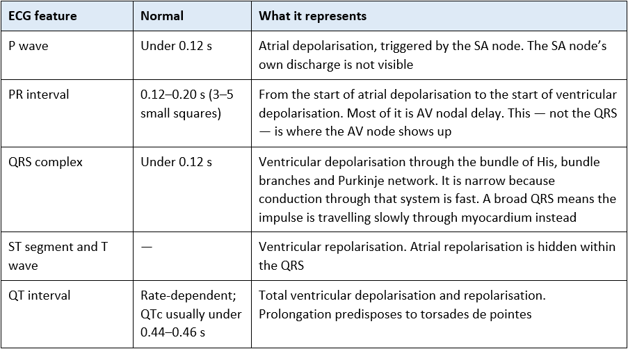
The reasoning shortcut
A long PR interval points to the AV node.
A broad QRS points to the conduction system below the AV node — the bundle branches or the ventricular myocardium itself.
An absent or dissociated P wave points to the SA node or to the atria.
Once you can place the problem on the pathway, the rhythm names follow.
See 12 lead ECG Essentials and Heart Rhythms – Overview.
8. When conduction fails: blocks and escape rhythms
8.1 Sinus node dysfunction
If the SA node slows or fails, a lower focus escapes. You will see sinus bradycardia, sinus pauses or arrest, or a junctional rhythm with absent, inverted or abnormally timed P waves. Always look for a reversible cause before assuming intrinsic nodal disease — see section 9.
8.2 Atrioventricular block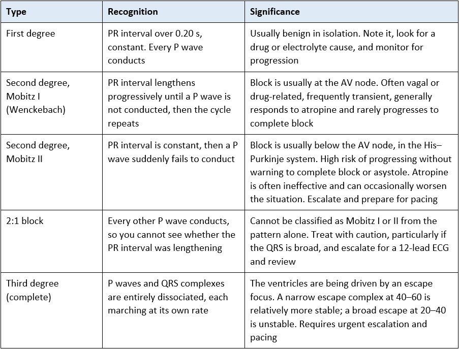
8.3 Bundle branch block
A QRS of 0.12 seconds or more indicates that one bundle branch is not conducting, so the ventricles depolarise sequentially rather than together. Left bundle branch block also makes the ECG difficult to interpret for ischaemia.
New left bundle branch block in a patient with ischaemic symptoms should be escalated urgently. Current guidance no longer treats new or presumed-new left bundle branch block as automatically equivalent to ST elevation; additional criteria are applied. Follow local and national acute coronary syndrome guidance — your job is to recognise the change and escalate it, not to classify it.
Safety point — broad complex tachycardia and accessory pathways
A regular broad complex tachycardia should be treated as ventricular tachycardia until proven otherwise.
An irregular broad complex tachycardia may be pre-excited atrial fibrillation, in which an accessory pathway bypasses the AV node. In this situation AV nodal blocking drugs — adenosine, verapamil, diltiazem, digoxin and beta blockers — can accelerate conduction down the accessory pathway and precipitate ventricular fibrillation.
Escalate immediately rather than reaching for standard rate control. Follow the Resuscitation Council UK tachycardia algorithm and local policy.
9. Reversible causes: electrolytes, drugs and stimuli
In critical care, most new conduction disturbance is secondary. Work through the reversible causes before concluding the problem is the conduction system itself.
9.1 Electrolytes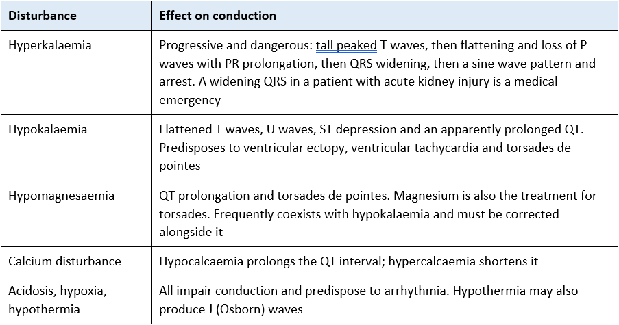
9.2 Drugs
Rate and AV conduction slowing: beta blockers, non-dihydropyridine calcium channel blockers (verapamil and diltiazem), digoxin, amiodarone, and alpha-2 agonists such as clonidine and dexmedetomidine. Propofol and opioids also commonly contribute.
Deliberate transient AV block: adenosine is used precisely because it blocks the AV node briefly — expect a short pause on the trace, warn the patient, and record a rhythm strip during administration.
QT prolongation: amiodarone, sotalol, haloperidol and other antipsychotics, ondansetron, macrolides, quinolones and some antifungals. Risk is cumulative when several are prescribed together, and is amplified by hypokalaemia and hypomagnesaemia. Check the QTc before adding another.
Perioperative and procedural: suxamethonium and neostigmine can cause bradycardia; suxamethonium also causes a transient potassium rise, which is dangerous in specific patient groups.
See Common Cardiac Drugs and Management of Cardiac Arrhythmias.
9.3 Other causes
– Ischaemia and infarction — see section 6 for the territory-specific pattern.
– Raised intracranial pressure — bradycardia with hypertension (Cushing’s reflex) is a neurological emergency, not a cardiac one.
– Recent cardiac surgery or transcatheter valve implantation — conduction tissue sits close to the aortic valve and the septum.
– Vagal stimuli — see section 5.
– Central line insertion — a guidewire or catheter tip in the right ventricle can provoke ectopy, and occasionally right bundle branch block or complete block in a patient who already has left bundle branch block.
10. Electrical activity is not mechanical output
Step 2 content — rationale and application
The monitor shows conduction, not contraction. A perfectly organised complex on the screen tells you the electrical system worked. It tells you nothing about whether the ventricle ejected any blood.
Pulseless electrical activity is organised electrical activity without a palpable pulse. The conduction system may be entirely normal; the problem is mechanical or circulatory. This is why cardiac arrest is confirmed by assessing the patient, not by reading the monitor — and why the reversible causes are the priority.
Always cross-check against the arterial trace where the patient has one. If the rhythm strip changes and the arterial waveform does not, suspect artefact. If the arterial waveform disappears and the rhythm strip does not, suspect the patient.
Before calling asystole, check the basics: leads attached, correct gain, correct lead selected. Fine ventricular fibrillation can look like asystole at low gain. Equally, movement, shivering, chest physiotherapy and filtration circuits all generate artefact that mimics arrhythmia.
Look at the patient. A patient sitting up talking to you is not in ventricular tachycardia, whatever the monitor says. This sounds obvious, and it is still the single most useful habit on the unit.
Waveform correlates you can use
Cannon a waves on the CVP trace or in the JVP occur when the atrium contracts against a closed tricuspid valve. They are seen in complete heart block, junctional rhythm and ventricular pacing — all situations where atrial and ventricular activity are dissociated. Intermittent large a waves are a useful clue that the patient is in complete heart block rather than sinus bradycardia.
Loss of the atrial contribution on the CVP or arterial trace often accompanies a fall in blood pressure when a patient moves from sinus rhythm into junctional rhythm, atrial fibrillation or ventricular pacing — see section 11.
11. Pacing: transcutaneous, temporary and permanent
Step 2 content — rationale and application
11.1 Why atrioventricular synchrony matters
Atrial contraction contributes the final 20 to 30% of ventricular filling — the atrial kick. In a normal compliant ventricle, losing it is tolerable. In a stiff, hypertrophied or poorly compliant ventricle — aortic stenosis, mitral stenosis, hypertrophic cardiomyopathy, diastolic dysfunction, or a ventricle after cardiac surgery — that contribution matters disproportionately.
This is the rationale behind pacing mode selection. Where the atria are working and the problem is conduction, a mode that preserves atrioventricular synchrony (for example atrial or dual-chamber pacing) will usually give a better blood pressure at the same rate than ventricular pacing alone. This is often visible at the bedside within a beat or two of a mode change — which is exactly the sort of observation a Step 2 assessor will ask you to explain.
11.2 Modalities
Transcutaneous pacing — an emergency bridge. It is painful, so the conscious patient needs analgesia and sedation. Confirm mechanical capture with a pulse or arterial trace — pacing spikes followed by a complex on the screen do not prove the heart ejected. Muscle twitching is not capture.
Temporary transvenous pacing — a wire placed via a central vein into the right ventricle. Watch for displacement, perforation, infection and failure to capture, and secure the wire and box carefully.
Epicardial pacing wires after cardiac surgery — sutured directly onto the heart and brought out through the chest wall. Confirm from the operation note which are atrial and which are ventricular before connecting anything. See Epicardial Pacing following Cardiac Surgery.
Permanent pacemakers and ICDs — know the device, the indication and whether the patient is pacing-dependent. Keep defibrillation pads at least 8 cm from the generator or use the anteroposterior position.
Pacing safety essentials
Check and document capture and sensing thresholds at the frequency your local policy requires, and after any change in the patient’s condition.
Know the three common failures: failure to capture (spike with no complex), failure to sense (spikes falling inappropriately, with the risk of an R-on-T event) and oversensing (inhibited pacing when it is needed).
Exposed pacing wires conduct current directly to the myocardium. Insulate the terminals, handle with gloves and follow local policy on microshock precautions.
Never leave a pacing-dependent patient without a backup plan and the equipment to deliver it. See Principles of Cardiac Pacing.
12. Clinical implications for the acute care nurse
Setting up monitoring
Choose a lead in which both the P wave and the QRS are clearly visible — lead II is usually the best starting point for P waves and gives you the most complete view across the heart.
Prepare the skin, site the electrodes correctly and change them when they degrade.
Set alarm limits for the individual patient rather than accepting defaults, and reset them when the clinical target changes.
Record and file a baseline rhythm strip, and a 12-lead ECG where indicated, so that later changes can be compared against something. See Cardiac Monitoring – Set Up.
Through the shift
Look at the trace, not just the number. A rate of 45 tells you very little; a rate of 45 with dissociated P waves and a broad QRS tells you a great deal.
Treat new conduction disturbance as a prompt to check potassium, magnesium, calcium, gases, temperature and the drug chart, escalate.
Anticipate vagal bradycardia around suctioning, intubation and NG insertion, and monitor throughout.
Document rhythm, rate, PR and QRS appearance and any change, and hand it over explicitly.
Escalate immediately for:
– Mobitz II or complete heart block, particularly with a broad escape complex or haemodynamic compromise — and bring the pacing equipment to the bedside while you escalate.
– New bundle branch block, especially with chest pain, after anterior infarction, or after cardiac surgery or TAVI.
– A widening QRS in a patient at risk of hyperkalaemia – this is a pre-arrest sign.
– Any broad complex tachycardia, and any irregular broad complex tachycardia without delay.
– Bradycardia with hypertension and a falling conscious level — think raised intracranial pressure, not primary cardiac disease.
– Failure to capture or failure to sense in any paced patient.
13. Self-test
Check your understanding. The section to check your answer against is shown in brackets.
Step 1
– Describe the normal conduction pathway in sequence, from the SA node to the ventricular myocardium. (Sections 2 and 3)
– Which part of the ECG complex corresponds to AV nodal delay, and what is its normal duration? (Section 7)
– State the intrinsic rate of the SA node, the AV junction and ventricular escape tissue. Why does the escape rate matter clinically? (Section 4)
– What does a QRS of 0.14 seconds tell you about where the impulse is travelling? (Section 7)
– How would you distinguish Mobitz I from Mobitz II on a rhythm strip, and which one worries you more? (Section 8.2)
– List six reversible causes of new bradycardia in a critically ill patient. (Section 9)
– Your patient’s monitor shows an organised rhythm at 70 but you cannot feel a pulse. What is this called and what do you do? (Section 10)
Step 2
– Explain why Mobitz I usually responds to atropine and Mobitz II usually does not, using the anatomical site of the block. (Sections 8.2 and 6)
– Your patient has an inferior STEMI and is bradycardic at 40 with first-degree block. Why is this anatomically predictable, and how does your concern differ from the same finding in an anterior STEMI? (Section 6)
– Your post-cardiac-surgery patient’s blood pressure falls when the pacing mode is changed from dual-chamber to ventricular-only at the same rate. Explain the mechanism. (Section 11.1)
– You notice intermittent very large a waves on the CVP trace in a bradycardic patient. What does this suggest and why? (Section 10)
– A patient on amiodarone and ondansetron has a potassium of 3.1 and a magnesium of 0.6. What are you worried about, and what would you check and correct? (Section 9)
– The transcutaneous pacer is showing spikes followed by wide complexes at 70, but the arterial trace is flat. -What has happened and what do you do? (Section 11.2)
– Why can AV nodal blocking drugs be dangerous in an irregular broad complex tachycardia? (Section 8.3)
Abbreviations
AF Atrial fibrillation
AV Atrioventricular
CC3N Critical Care Networks – National Nurse Leads
CVP Central venous pressure
ECG Electrocardiogram
ICD Implantable cardioverter defibrillator
ICP Intracranial pressure
JVP Jugular venous pressure
LAD Left anterior descending artery
LBBB / RBBB Left / right bundle branch block
PEA Pulseless electrical activity
QTc Corrected QT interval
SA Sinoatrial
STEMI ST elevation myocardial infarction
TAVI Transcatheter aortic valve implantation
VF / VT Ventricular fibrillation / ventricular tachycardia
Glossary
Automaticity — the ability of cardiac tissue to depolarise spontaneously without an external stimulus.
Capture — a paced impulse successfully producing depolarisation and, ideally, contraction.
Escape rhythm — a rhythm arising from a lower pacemaker when the one above it fails or is blocked.
AV dissociation — atria and ventricles depolarising independently of one another.
Infranodal — below the AV node, that is within the His–Purkinje system.
Pre-excitation — early ventricular activation via an accessory pathway that bypasses the AV node.
Sensing — a pacemaker’s detection of the patient’s own intrinsic beats, so that it does not pace unnecessarily.
Torsades de pointes — a polymorphic ventricular tachycardia associated with QT prolongation.
Recommended pages
Next in this section
Cardiac Action Potential — the cellular basis of automaticity and conduction
The Cardiac Cycle — how electrical events translate into mechanical ones
Heart Valves — the insulating role of the fibrous skeleton and the atrial kick
Cardiac Surface Anatomy — electrode and pad placement landmarks
Applying this clinically
Heart Rhythms – Overview; Management of Cardiac Arrhythmias
12 lead ECG Placement; 12 lead ECG Essentials; 12 lead ECG territories
Cardiac Monitoring – Set Up
Principles of Cardiac Pacing; Epicardial Pacing following Cardiac Surgery
Coronary Artery Anatomy — blood supply to the conduction system
Common Cardiac Drugs
Acute Kidney Injury — hyperkalaemia and its ECG changes
Traumatic Brain Injury Management – ICU — Cushing’s reflex
Further reading and local governance
Guidelines
Resuscitation Council UK — adult advanced life support, and the peri-arrest bradycardia and tachycardia algorithms.
ESC Guidelines on cardiac pacing and cardiac resynchronisation therapy.
ESC Guidelines for the management of patients with ventricular arrhythmias and the prevention of sudden cardiac death.
NICE NG196 — Atrial fibrillation: diagnosis and management.
UK Renal Association / national guidance on the treatment of acute hyperkalaemia in adults.
CC3N National Competency Framework for Registered Nurses in Adult Critical Care — Step 1 (1:3.7) and Step 2 (2:2.1).
Local governance
Your local peri-arrest, cardiac arrest and escalation policies.
Your local temporary and epicardial pacing policy, including threshold checking frequency and microshock precautions.
Your local hyperkalaemia and electrolyte replacement protocols.
Your local guidance on QT monitoring and high-risk drug combinations.
Textbooks
Levick, An Introduction to Cardiovascular Physiology
Adam, Osborne and Welch, Critical Care Nursing: Science and Practice (Oxford)
Bersten and Handy, Oh’s Intensive Care Manual
Last reviewed: July 2026 Next review due: July 2027. AskAlth Nursing Team, London UK.
AskAlth is an educational resource for registered healthcare professionals in UK adult critical care. Not for patients or the public. Not a substitute for local policy, clinical guidance or the BNF. Content reflects guidance at the time of writing. AskAlth is independent and not affiliated with CC3N, NICE, the Resuscitation Council UK, the NMC or the NHS.
AskAlth is an independent educational resource created by NHS doctors and critical care nurses. It is not affiliated with CC3N, NICE, or the NHS, though our content is mapped to and referenced against their published frameworks and guidance.