Coronary Artery Structure and Histology
Contents
How to use this resource, and learning outcomes
1. Why the vessel wall matters at the bedside
2. The three layers of the coronary artery wall
3. The endothelium is an organ, not a lining
4. What makes the coronary arteries different
5. Histological appearance under the microscope
6. What goes wrong: from endothelial dysfunction to plaque rupture
7. Clinical implications for the acute care nurse
8. Test your knowledge
Abbreviations
Further reading and local governance
How to use this resource
This page sits alongside Coronary Artery Anatomy. That resource tells you where the arteries run and what they supply; this one tells you what they are made of, and why that matters when a patient in front of you has chest pain, a stent, or an infarct.
If you are new to the topic, read sections 2 and 3 for the structure, then go to sections 6 and 7 for the clinical link.
Learning outcomes
By the end of this session you will be able to:
– Name the three layers of the coronary artery wall and describe the tissue in each.
– Explain why the coronary arteries are classed as muscular rather than elastic arteries.
– Describe at least three functions of the vascular endothelium.
– Explain how atherosclerosis develops within the intima, and why plaque rupture causes acute coronary syndrome.
– Relate the structure of the vessel wall to the actions of nitrates, antiplatelets and coronary stents.
1. Why the vessel wall matters at the bedside
It is tempting to treat histology as exam content that stops being relevant once you are on the unit. It does not. Almost everything you do for a cardiac patient acts on one of these three layers.
Atherosclerosis is a disease of the intima. It begins with injury to a single layer of cells, decades before the patient presents.
Vasospasm and vasodilation are functions of the media. Glyceryl trinitrate works by relaxing the smooth muscle in that layer.
Plaque rupture is a structural failure. The acute coronary syndromes are what happens when the cap over a plaque tears and exposes the material underneath to flowing blood.
Stents work, and fail, at the level of the vessel wall. Restenosis and stent thrombosis are both consequences of how the wall heals.
2. The three layers of the coronary artery wall
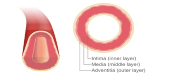
The coronary arteries are muscular (medium-sized) arteries. They follow the standard three-layer arterial plan, but with features that reflect their workload: they sit on the surface of a moving organ, are compressed with every systole, and are exposed to constant pulsatile pressure.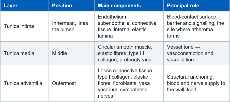
2.1 Tunica intima
Endothelium — a single layer of simple squamous endothelial cells in direct contact with the blood.
Subendothelial layer — loose connective tissue with collagen, elastic fibres and occasional smooth muscle cells.
Internal elastic lamina (IEL) — a well-defined, fenestrated sheet of elastin marking the boundary with the media. The fenestrations allow nutrients to diffuse inwards from the lumen.
The coronary arteries characteristically develop diffuse intimal thickening with age. A degree of this is a normal finding rather than disease, but it is also the soil in which atheroma develops.
2.2 Tunica media
Predominantly circularly arranged smooth muscle cells, which is what defines a muscular artery.
Also contains elastic fibres, type III collagen and proteoglycans.
Elastic tissue content sits between the two extremes: less than an elastic artery such as the aorta, more than a small muscular artery. The aorta needs elastin to store energy in systole and recoil in diastole; the coronaries need muscle to regulate their own diameter.
An external elastic lamina (EEL) marks the boundary with the adventitia, though it is less distinct than the IEL.
In the larger coronary arteries, vasa vasorum reach the outer media. The inner wall is nourished by diffusion from the lumen.
2.3 Tunica adventitia
Loose connective tissue, type I collagen, elastic fibres and fibroblasts.
Carries the vasa vasorum and sympathetic nerve fibres.
Blends with the surrounding epicardial adipose tissue, since the major coronary arteries run over the surface of the heart within that fat before their branches penetrate the myocardium.
3. The endothelium is an organ, not a lining
This is the single most useful thing to take from this resource. The endothelium is metabolically active and continuously regulates the vessel beneath it.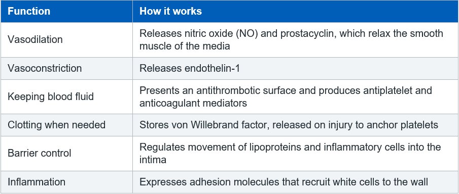

4. What makes the coronary arteries different
Four features distinguish them from other muscular arteries of similar size.
a. They are compressed during systole. Perfusion is therefore mainly diastolic, particularly on the left. This is covered in the Coronary Artery Anatomy resource.
b. They move constantly. The epicardial vessels flex with every beat, producing repetitive mechanical stress that favours intimal injury, particularly at branch points and bends where blood flow becomes turbulent.
c. They are conduit vessels. The epicardial arteries seen on an angiogram contribute little to resistance. Resistance and autoregulation sit in the intramyocardial arterioles downstream. This is why a stenosis often causes no symptoms until it exceeds roughly 70% of the lumen — the arterioles dilate to compensate, using up the coronary flow reserve. Once that reserve is exhausted, symptoms appear.
d. Myocardial bridging. In some people a segment of artery, most often the LAD, tunnels through the myocardium rather than running over it, and is squeezed with each systole.
5. Histological appearance under the microscope
On a standard transverse section:
Endothelium lining a relatively narrow lumen, often thrown into folds if the vessel was fixed in a contracted state.
Internal elastic lamina as a distinct wavy line beneath the intima — usually the easiest landmark for orientating yourself on the slide.
Concentric layers of smooth muscle in the media, with elastic fibres demonstrated using elastic stains such as Verhoeff–Van Gieson or Weigert’s elastic stain. On a routine haematoxylin and eosin section the elastic fibres are not well shown.
External elastic lamina at the media–adventitia border, less distinct than the IEL.
Vasa vasorum and nerve fibres within the adventitia, with adipose tissue beyond.
Orientation tip
On the same slide, the accompanying coronary vein has a much thinner wall, a larger and more irregular lumen, and no well-defined internal elastic lamina.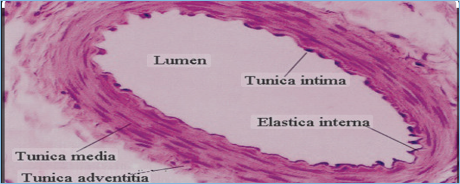
6. What goes wrong: from endothelial dysfunction to plaque rupture
Atherosclerosis is an inflammatory process that takes place within the intima.
Endothelial injury and dysfunction. Risk factors and turbulent flow at branch points damage the endothelium. Its permeability rises and it becomes pro-inflammatory.
Lipid entry and oxidation. Low-density lipoprotein passes into the subendothelial space and is oxidised.
Inflammatory cell recruitment. Monocytes adhere, migrate in, become macrophages and take up the oxidised lipid to form foam cells. Visible at this stage as a fatty streak.
Plaque formation. Smooth muscle cells migrate from the media into the intima and lay down collagen, forming a fibrous cap over a lipid-rich necrotic core.
Plaque instability. Ongoing inflammation thins the cap. Vasa vasorum grow into the plaque and may bleed, expanding it from within.
Rupture and thrombosis. The cap tears, exposing collagen and tissue factor to flowing blood. Platelets adhere and aggregate, and a thrombus forms. This is the event behind unstable angina, NSTEMI and STEMI.
Two points that surprise people
(i) The plaque that ruptures is often not the tightest one. A soft, lipid-rich, thin-capped plaque causing 40% stenosis is more dangerous than a heavily calcified one causing 80%. This is why a patient can have a normal exercise test and arrest a fortnight later.
(ii) Plaques can grow outwards before they grow inwards. The vessel initially remodels to preserve the lumen, so significant disease can be present before an angiogram shows narrowing.
7. Clinical implications for the acute care nurse
Nitrates act on the media
Glyceryl trinitrate is a nitric oxide donor. It supplies NO directly to the smooth muscle, which is why it still works when the endothelium is too diseased to produce its own. Its main antianginal benefit is venodilation and reduced preload rather than coronary dilation.
Antiplatelets act on the intima
Aspirin and P2Y12 inhibitors target the platelet response to exposed subendothelial collagen. This is why dual antiplatelet therapy is not something to omit casually, and why interrupting it after recent stenting must be a senior decision.
Stents are a wall-healing problem
A stent injures the intima by design.
In-stent restenosis is neointimal hyperplasia — smooth muscle proliferating into the lumen over months. Drug-eluting stents release agents that suppress this.
Stent thrombosis happens when the metal is not yet covered by endothelium and is exposed to blood. It is sudden, often presents as a STEMI, and is strongly linked to stopping antiplatelet therapy early. Recurrent chest pain or ST re-elevation after PCI needs immediate escalation.
Coronary spasm can occur with normal arteries
Intense contraction of the media causes ischaemia without a fixed stenosis, classically in variant (Prinzmetal) angina. Cocaine is an important cause in acute presentations. In suspected cocaine-associated chest pain, beta blockers are generally avoided and benzodiazepines with nitrates are usually preferred. Follow your local protocol.
Risk factor control is endothelial protection
When you deliver smoking cessation advice, glycaemic control or a statin, you are acting on the layer where the disease starts. Statins improve endothelial function as well as lowering lipids.
8. Test your knowledge
Complete these before your competency discussion.
– Which layer of the artery wall does atherosclerosis develop in?
– Name two vasoactive substances produced by the endothelium and state the effect of each.
– Why are the coronary arteries classed as muscular rather than elastic arteries?
_ A patient’s angiogram shows a 45% stenosis. Why does this not mean they are at low risk of an acute event?
Explain, in terms of the vessel wall, why stopping dual antiplatelet therapy early after stent insertion is dangerous.
_ Which stain would you request to demonstrate the elastic laminae, and why will a routine H&E section not show them well?
Abbreviations
DAPT Dual antiplatelet therapy
EEL External elastic lamina
GTN Glyceryl trinitrate
H&E Haematoxylin and eosin
IEL Internal elastic lamina
LAD Left anterior descending artery
LDL Low-density lipoprotein
NO Nitric oxide
NSTEMI Non-ST-elevation myocardial infarction
PCI Percutaneous coronary intervention
STEMI ST-elevation myocardial infarction
Related pages:
Before this page:
Coronary Artery Anatomy — where these vessels run and what they supply
Structure and Function of the Heart — chambers, walls and layers
The Cardiac Cycle — why these arteries are compressed in systole
Alongside this page:
Coronary Veins — thinner walls, no internal elastic lamina
Cardiac v Skeletal Muscle — the tissue these arteries supply
Cardiac Surface Anatomy — the epicardial fat the adventitia blends into
When the wall fails:
Coronary Artery Disease — atheroma, stenosis and flow reserve
Stable Angina — a fixed stenosis limiting flow on exertion
Unstable Angina — plaque instability without infarction
Acute Coronary Syndrome — what plaque rupture looks like clinically
Myocardial Infarction — occlusion, territory and complications
Hypertension — chronic pressure loading of the arterial wall
Common Cardiac Drugs — nitrates, antiplatelets and statins, and where each acts
Working through your competencies? Follow the Step 1 ICU Pathway.
Further reading and local governance
CC3N National Competency Framework for Registered Nurses in Adult Critical Care, Step 1 — cardiovascular section.
Your local acute coronary syndrome and antiplatelet guidelines, including advice on interrupting dual antiplatelet therapy.
NICE guidance on acute coronary syndromes and on lipid modification.
A current histology or cardiovascular pathology text for the underpinning science.
Note for the practice educator
This document supports competency development but does not replace assessment in practice.
All clinical management points, particularly those relating to antiplatelet therapy and cocaine-associated chest pain, must be read alongside current local and national guidance and reviewed against them before use.
AskAlth is an independent educational resource created by NHS critical care nurses. It is not affiliated with CC3N, NICE, or the NHS, though our content is mapped to and referenced against their published frameworks and guidance.