

Cardiac structure and chambers
The human heart is a muscular organ that functions as the central pump of the circulatory system, maintaining the continuous flow of blood throughout the body to supply oxygen and nutrients while removing waste products. Structurally, the heart is divided into four chambers—two atria and two ventricles—that, in normal function, work together in a coordinated manner to ensure efficient circulation.
External Structure
The heart is enclosed within a protective double-walled sac known as the pericardium, which contains pericardial fluid to reduce friction during contractions. The outer layer of the heart wall is the epicardium, the middle layer is the myocardium (composed of cardiac muscle responsible for pumping action), and the inner layer is the endocardium, which lines the chambers and valves.
Externally, the heart shows a few visible features such as the auricles—small flap-like projections on the atria that increase their capacity—and several grooves or sulci that mark the boundaries between chambers and house the major coronary blood vessels. The coronary arteries supply oxygenated blood to the heart muscle itself, while the cardiac veins drain deoxygenated blood into the coronary sinus.
Internal Structure and Chambers
Internally, the heart is divided into four chambers: the right atrium, right ventricle, left atrium, and left ventricle. A muscular wall called the septum separates the right and left sides, preventing the mixing of oxygenated and deoxygenated blood. 
Right Atrium:
The right atrium receives deoxygenated blood from the body through three major veins—the superior vena cava, inferior vena cava, and coronary sinus.
It acts as a receiving chamber, holding blood temporarily before it passes through the tricuspid valve into the right ventricle. The Tricuspid valve ensures one-way flow (from the right atrium to the right ventricle), preventing backflow during ventricular contraction (systole).
Right Ventricle:
The right ventricle pumps deoxygenated blood to the lungs for oxygenation through the pulmonary artery. The flow is regulated by the pulmonary valve, which opens during contraction and closes to prevent backflow when the ventricle relaxes. The muscular wall of the right ventricle is thinner than the left because it only needs to pump blood to the nearby lungs under low pressure.
Left Atrium:
The left atrium receives oxygenated blood from the lungs via four pulmonary veins. It then pushes the blood through the mitral valve into the left ventricle. Like the right atrium, its wall is thin since it only needs to move blood a short distance.
Left Ventricle:
The left ventricle has the thickest muscular wall because it must generate enough force to pump blood through the aorta to the entire body, during systole. The aortic valve regulates blood flow from the left ventricle into the aorta and prevents backflow during relaxation (diastole).
Blood Flow Through the Heart
Blood moves through two circuits in series — the pulmonary circulation (right heart to lungs) and the systemic circulation (left heart to body) — with the heart acting as a double pump.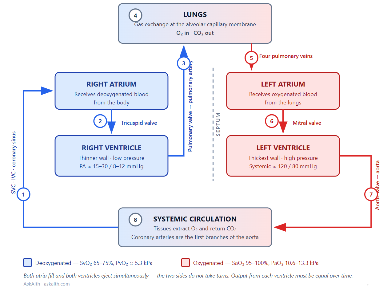
Pulmonary circuit (right side):
Deoxygenated blood enters the right atrium from the superior vena cava (head, neck, upper limbs), inferior vena cava (abdomen, pelvis, lower limbs) and coronary sinus (heart muscle itself) (1).
It passes through the tricuspid valve into the right ventricle (2).
Right ventricular contraction opens the pulmonary valve, ejecting blood into the pulmonary trunk, which divides into the left and right pulmonary arteries (3).
Note: Artery and vein describe direction, not oxygen content. Arteries carry blood away from the heart; veins carry it towards the heart. The pulmonary arteries are the only arteries carrying deoxygenated blood, and the pulmonary veins the only veins carrying oxygenated blood.
At the alveolar capillary membrane, gas exchange occurs: oxygen diffuses into the blood, carbon dioxide diffuses out (4).
Systemic circuit (left side):
Now oxygenated, blood returns via four pulmonary veins into the left atrium (5).
It passes through the mitral valve into the left ventricle (6).
Left ventricular contraction opens the aortic valve, ejecting blood into the aorta and on to the systemic circulation — including the coronary arteries, which are the first branches off the aortic root (7).
Having delivered oxygen and collected carbon dioxide at the tissues (8), blood returns to the right atrium and the cycle repeats (1).
A common misconception: the two sides of the heart do not take turns. Both atria fill and both ventricles eject simultaneously. Over time, output from the right and left ventricles must be equal — a sustained mismatch of even a few percent would rapidly cause pulmonary or systemic congestion.
What differs is pressure, not volume.
The right ventricle ejects into a low-resistance pulmonary circuit (pulmonary artery pressure typically 15–30/8–12 mmHg), while the left ejects against high systemic resistance (around 120/80 mmHg). This is why the left ventricular wall is roughly three times thicker despite handling identical stroke volume.
Oxygenated and Deoxygenated Blood.
The terms “oxygenated” and “deoxygenated” describe the oxygen content of blood at a given point in the circuit, not two different types of blood.
Deoxygenated blood returns from the tissues having given up oxygen and picked up carbon dioxide. In a healthy adult it has a mixed venous oxygen saturation (SvO₂) of roughly 65–75%, a PvO₂ around 5.3 kPa (40 mmHg), and a PvCO₂ around 6.1 kPa (46 mmHg). It occupies the right side of the heart and the pulmonary arteries.
Oxygenated blood has passed through the pulmonary capillaries and equilibrated with alveolar gas. Arterial saturation (SaO₂) is normally 95–100%, with a PaO₂ of 10.6–13.3 kPa (80–100 mmHg) and a PaCO₂ of 4.6–6.0 kPa. It occupies the left side of the heart and the systemic arteries.
Why this matters clinically
The septum normally keeps the two circuits entirely separate. Where it doesn’t — VSD, ASD, patent foramen ovale — blood shunts between circuits. A right-to-left shunt allows deoxygenated blood to bypass the lungs entirely, producing hypoxaemia that does not correct with supplemental oxygen. This is a key differentiator from V/Q mismatch.
ScvO₂ (from a central line, tip in SVC/RA) and SvO₂ (from a pulmonary artery catheter) sample deoxygenated blood before it reaches the lungs. A falling value suggests the tissues are extracting more oxygen than usual — typically low cardiac output, anaemia, hypoxaemia, or raised demand.
Arterial blood gases must be taken from an artery, after the lungs, because venous sampling reflects tissue extraction rather than pulmonary gas exchange.
Understanding the sequence is what makes valve lesions intuitive: mitral stenosis backs pressure up into the pulmonary veins; tricuspid regurgitation backs it up into the systemic veins and produces the raised JVP and peripheral oedema you see on the unit.
The heart’s four-chambered structure allows for the complete separation of oxygenated and deoxygenated blood, a key adaptation for efficient double circulation in humans. This design ensures that oxygen-rich blood is delivered to tissues at high pressure, while deoxygenated blood is sent to the lungs for gas exchange at lower pressure. Through its rhythmic contractions and coordinated valve system, the heart maintains a precise, unidirectional flow that sustains life.
Related AskAlth Pages
Next in the anatomy sequence
Heart Valves — the four valves in detail, and what happens when they fail
The Cardiac Cycle — the timing of filling and ejection described above
Cardiac Surface Anatomy
Coronary Artery Anatomy and Coronary Veins — the heart’s own blood supply and drainage into the coronary sinus
Starling’s Law — why right and left ventricular output stay matched
Where this shows up at the bedside
Pulse Oximetry — measuring oxygen saturations
Arterial Blood Gas – Overview — why the sample must be arterial
V/Q Mismatch — distinguishing shunt from mismatch
Cardiac Output Monitoring and Cardiac Output Studies — including SvO₂ and ScvO₂ interpretation
Invasive Monitoring – Waveforms (Art, CVP, PA, PCWP) — pressures through each chamber
Normal observation values
When the circuits fail
Left Ventricular Failure — backward pressure into the pulmonary circulation
Cor Pulmonale — right heart failure secondary to lung disease
Pathophysiology of Heart Failure
Cardiogenic Shock
Pulmonary Embolism (PE) — obstruction of the pulmonary circuit
Acquired Heart Valve Disease
Further Reading
Open access
Rehman I, Rehman A. Anatomy, Thorax, Heart. StatPearls, updated 2023 — the core chamber-by-chamber reference on NCBI Bookshelf NCBI
Tucker WD, Weber C, Burns B. Anatomy, Thorax, Heart Pulmonary Arteries. StatPearls, 2023
Shahoud JS, Miao JH, Bolla SR. Anatomy, Thorax, Heart Aorta. StatPearls, 2023
Bamalan OA, Jozsa F, Soos MP. Anatomy, Thorax, Heart Great Vessels. StatPearls, 2023 — covers the venae cavae, pulmonary vessels and aortic root as a group, and how they carry blood to the lungs and back out to the periphery NCBI
Textbooks
Marino PL. The ICU Book / Marino’s The Little ICU Book — the standard reference for oxygen delivery, extraction and venous saturation interpretation
Bersten AD, Handy JM (eds). Oh’s Intensive Care Manual
Levick JR. An Introduction to Cardiovascular Physiology
Tortora GJ, Derrickson B. Principles of Anatomy and Physiology — for the underlying A&P if it’s been a while
Specialist
BJA Education has accessible review articles on pulmonary circulation, right ventricular function and shunt physiology — searchable free on ScienceDirect
Last Updated, AskAlth Nursing Team, July 2026, London UK
AskAlth is an independent educational resource created by NHS doctors and critical care nurses. It is not affiliated with CC3N, NICE, or the NHS, though our content is mapped to and referenced against their published frameworks and guidance.