

Coronary Veins
Contents
How to use this resource, and learning outcomes
1. Why venous drainage matters at the bedside
2. Three routes back to the right atrium
3. The coronary sinus
4. Tributaries of the coronary sinus
5. The anterior cardiac veins
6. The smallest cardiac veins (Thebesian veins)
7. Clinical implications for the acute care nurse
8. Test your knowledge
Abbreviations
Related Pages
Further reading and local governance
How to use this resource
This is the counterpart to Coronary Artery Anatomy. The arteries deliver; these vessels take blood away. The venous side is usually taught quickly and forgotten, but it is the route used for biventricular pacing leads and retrograde cardioplegia, and it explains why arterial oxygen saturation is never quite 100%.
Learning outcomes
– By the end of this session you will be able to:
– Describe the position of the coronary sinus and where it drains.
– Name the main tributaries of the coronary sinus and state the region each drains.
– Explain the three separate routes by which venous blood returns from the myocardium.
– Explain why the Thebesian veins contribute to normal physiological shunt.
– Describe the clinical relevance of the coronary sinus to cardiac resynchronisation therapy, and the significance of a dilated coronary sinus on echocardiography.
1. Why venous drainage matters at the bedside
Three reasons this is worth knowing properly rather than skimming.
The coronary sinus is an access route. The left ventricular lead of a biventricular pacemaker is passed into it and out along one of its tributaries. Retrograde cardioplegia in cardiac surgery is delivered through it.
It is a landmark for the conduction system. The opening of the coronary sinus forms one border of the triangle of Koch, which contains the atrioventricular node.
Some venous blood bypasses the lungs entirely. The smallest cardiac veins empty directly into the heart chambers, adding a small volume of deoxygenated blood to the arterial side. This is part of the normal physiological shunt.
2. Three routes back to the right atrium
Venous blood leaves the myocardium by three separate routes, not one. Most textbook diagrams show only the first.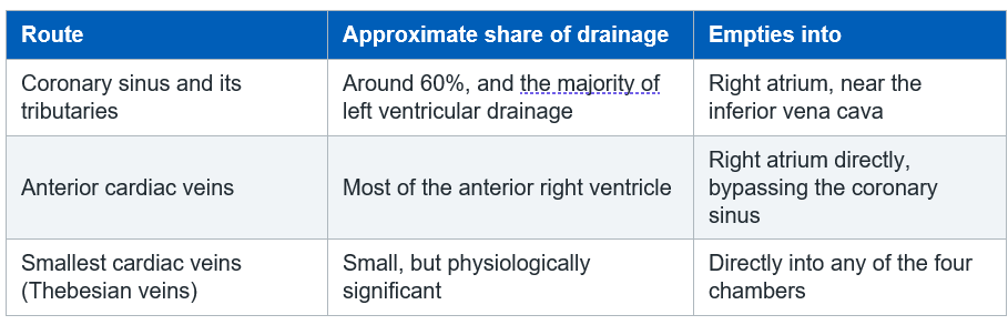
Proportions vary between sources and between individuals. Use them as approximations.
3. The coronary sinus
The largest vein of the heart.
Position: lies in the posterior part of the coronary sulcus (the atrioventricular groove), between the left atrium and the left ventricle, on the posterior surface of the heart.
Origin: it is the continuation of the great cardiac vein. The transition is conventionally marked by the point at which the oblique vein of the left atrium joins, and by the valve of Vieussens.
Termination: opens into the right atrium, between the opening of the inferior vena cava and the tricuspid valve orifice. The opening is partially guarded by the Thebesian valve.
Receives: the great, middle, small, posterior and oblique veins.
Landmark point
The coronary sinus ostium, the tendon of Todaro and the septal leaflet of the tricuspid valve form the boundaries of the triangle of Koch, within which the atrioventricular node sits.
This is why the region is handled cautiously during pacing lead placement and ablation.
4. Tributaries of the coronary sinus
A note on wording
Veins drain a territory; arteries supply it. Each vein below drains the region supplied by the artery it accompanies.
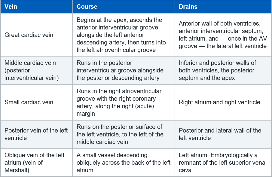
The posterior and lateral tributaries matter clinically: these are the vessels a cardiac physiologist is trying to reach when placing a left ventricular pacing lead, because pacing the lateral or posterolateral left ventricular wall is what achieves resynchronisation.
5. The anterior cardiac veins
Arise from the anterior surface of the right ventricle.
Cross the right atrioventricular groove.
Drain directly into the right atrium, bypassing the coronary sinus altogether. Some join the small cardiac vein first.
This route is the one most often left out of diagrams, and it accounts for most of the drainage of the right ventricle.
6. The smallest cardiac veins (Thebesian veins)
Tiny venous channels within the myocardial wall.
Open directly into the chambers of the heart, most numerously into the right atrium and right ventricle, but also into the left-sided chambers.
Why this matters physiologically
Thebesian veins draining into the left atrium and left ventricle add a small amount of deoxygenated blood to blood that has already passed through the lungs.
Together with the bronchial circulation, this is the normal anatomical (physiological) shunt. It is why a healthy person breathing room air has an arterial oxygen saturation in the high nineties rather than 100%, and a small but normal alveolar-arterial gradient.
7. Clinical implications for the acute care nurse
Cardiac resynchronisation therapy (CRT)
The left ventricular lead is not placed inside the left ventricle. It is advanced through the coronary sinus and wedged into a lateral or posterolateral tributary, pacing the left ventricle from the epicardial surface.
Complications you may see afterwards include coronary sinus dissection or perforation, lead displacement with loss of left ventricular capture, and phrenic nerve stimulation causing persistent hiccups or diaphragmatic twitching. Report any of these.
A dilated coronary sinus on echocardiography
Two common explanations, and both influence your practice.
Raised right atrial pressure, from right heart failure, pulmonary hypertension or tricuspid regurgitation.
A persistent left superior vena cava, which drains into the coronary sinus. This is the commonest thoracic venous anomaly. It matters when a central line is placed from the left side, because the catheter can follow this route and its tip will sit in the coronary sinus rather than the superior vena cava. Escalate an unexpected left-sided catheter course or a chest film that does not look right rather than simply using the line.
Cardiac surgery
Retrograde cardioplegia is delivered through a catheter placed in the coronary sinus, allowing the solution to perfuse the myocardium backwards through the venous system. It is used where antegrade delivery through the coronary arteries would be unreliable, such as in severe coronary disease or aortic regurgitation.
Electrophysiology
A catheter is routinely positioned in the coronary sinus during ablation procedures, both to record left atrial activity and as an anatomical reference. Chest pain with haemodynamic instability afterwards should raise the possibility of perforation and tamponade.
Coronary sinus rhythm
An ectopic atrial rhythm arising near the coronary sinus produces inverted P waves in the inferior leads with a normal or short PR interval. It is usually benign, but it is a recognised source of confusion on a monitor.
8. Test your knowledge
– Where does the coronary sinus lie, and where does it open?
– Which vein runs alongside the left anterior descending artery, and what does it drain?
– Name the three routes by which venous blood returns from the myocardium.
– Why does a healthy person breathing room air not achieve an arterial oxygen saturation of 100%?
– A patient is admitted after CRT implantation with persistent hiccups. What is the likely explanation, and what would you do?
– An echocardiogram reports a dilated coronary sinus. Give two possible causes.
Abbreviations
AV Atrioventricular
CRT Cardiac resynchronisation therapy
IVC Inferior vena cava
LAD Left anterior descending artery
LV Left ventricle
PDA Posterior descending artery
RA Right atrium
RV Right ventricle
SVC Superior vena cava Source: Radiopedia.
Related pages:
Before this page:
Coronary Artery Anatomy — the arterial side, and the grooves these veins share
Cardiac Surface Anatomy — the sulci, the crux and the posterior surface
Structure and Function of the Heart — chambers, walls and layers
Alongside this page
Coronary Artery Structure and Histology — why a vein wall looks so different down the microscope
Cardiac Conduction System — the AV node sits in the triangle of Koch, bordered by the coronary sinus
The Cardiac Cycle — filling, emptying and venous return
Apply it at the bedside:
Principles of Cardiac Pacing — the LV lead is placed through the coronary sinus
Invasive Monitoring – Waveforms — right atrial pressure and central line position
Heart Rhythms – Overview — including coronary sinus rhythm
Myocardial Infarction — the territories these veins drain
Left Ventricular Failure — the group most likely to receive resynchronisation therapy
Working through your competencies? Follow the Step 1 ICU Pathway.
Further reading and local governance
CC3N National Competency Framework for Registered Nurses in Adult Critical Care, Step 1 — cardiovascular section.
Your local guidance on central venous catheter insertion and confirmation of tip position.
Your local cardiac device and post-implantation care policy.
A current anatomy text for the underpinning anatomy.
AskAlth is an independent educational resource created by NHS critical care nurses. It is not affiliated with CC3N, NICE, or the NHS, though our content is mapped to and referenced against their published frameworks and guidance.