

Cardiac Myocyte Action Potential
How to use this resource, and learning outcomes:
1. Why the action potential matters at the bedside
2. The resting membrane potential
3. The ventricular (fast response) action potential
4. The pacemaker (slow response) action potential
5. Refractory periods and why they protect the heart
6. Excitation–contraction coupling: from calcium to squeeze
7. Linking the action potential to the ECG
8. Electrolytes and the action potential
9. Drugs and the action potential
10. Ischaemia, hypoxia and acidosis
11. Clinical implications for the acute care nurse
Abbreviations
12. Self-test
Glossary
Recommended pages
Further reading and local governance
How to use this resource
Sections 2 to 6 build the cellular physiology. Sections 7 to 11 apply it — to the ECG in front of you, to the potassium result you have just chased, to the infusion you are titrating and to the drug chart you are checking.
ICU Step Competencies:
If you are working through Step 1, sections 2 to 5, 7 and 11 are your priority — and section 8, because hyperkalaemia is named explicitly in the framework.
If you are working through Step 2 or an academic critical care programme, sections 6, 9 and 10 are where the rationale sits: this is the page that lets you explain why a drug or an electrolyte does what it does, rather than only that it does.
Learning outcomes
Step 1 — by the end of this session you will be able to:
– Describe the five phases of the ventricular action potential, naming the principal ion movement in each.
– Explain what maintains the resting membrane potential and which ion determines it.
– Describe how the pacemaker action potential differs from the ventricular one, and why that difference matters.
– Explain what the refractory period is and why it prevents the heart from going into tetany.
– State which part of the ECG corresponds to which phase of the action potential.
– Describe the effect of hyperkalaemia and hypokalaemia on the action potential and on the ECG.
Step 2 — in addition, you will be able to:
– Explain excitation–contraction coupling and use it to give a rationale for the action of beta agonists, phosphodiesterase inhibitors and digoxin.
– Map the Vaughan Williams antiarrhythmic classes onto the phases of the action potential.
– Explain why calcium is given in hyperkalaemia even though it does not lower the potassium, and why insulin, dextrose and nebulised salbutamol do something different.
– Explain why hypokalaemia will not correct until magnesium is replaced, and why hypokalaemia increases digoxin toxicity at any given level.
– Explain, at cellular level, why ischaemia and reperfusion are arrhythmogenic.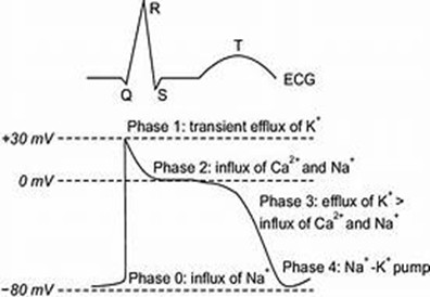
1. Why the action potential matters at the bedside
Cardiac myocytes have a distinctive action potential with a prolonged plateau phase, which is what allows the heart to contract effectively and then refill. The ventricular action potential lasts roughly 200 to 400 milliseconds — far longer than a nerve or skeletal muscle action potential.
This is not background theory. It is the level at which almost everything you do to the cardiovascular system actually works. Every antiarrhythmic, every inotrope, every potassium and magnesium result, every defibrillation and every ischaemic ECG change is an event happening on this trace.
Three questions this page answers: why a potassium of 7.1 widens the QRS; why calcium is given in hyperkalaemia even though it does not remove any potassium; and why a shock delivered on the T wave can kill someone while the same shock 100 milliseconds earlier is harmless.
2. The resting membrane potential
A resting ventricular myocyte sits at approximately −90 mV — the inside of the cell is negative relative to the outside.
Potassium sets it. At rest the membrane is far more permeable to potassium than to any other ion, through inward rectifier channels (IK1). The resting potential therefore sits close to the equilibrium potential for potassium, and is determined mainly by the ratio of potassium inside the cell to potassium outside it.
The sodium–potassium ATPase maintains the gradients. It pumps three sodium ions out for every two potassium ions in, using ATP. It is the reason the gradients do not simply run down, and it is the target of digoxin.
The single most useful consequence
Because the resting potential depends on the potassium ratio across the membrane, and because the extracellular potassium is the small number in that ratio, a change in serum potassium has a disproportionate effect.
Raise extracellular potassium and the cell becomes less negative at rest — partially depolarised. Lower it and the cell becomes more negative — hyper-polarised.
Everything in section 8 follows from that one sentence.
3. The ventricular (fast response) action potential
This is the action potential of working atrial and ventricular myocytes and of the His–Purkinje system — tissue whose job is to conduct fast and contract.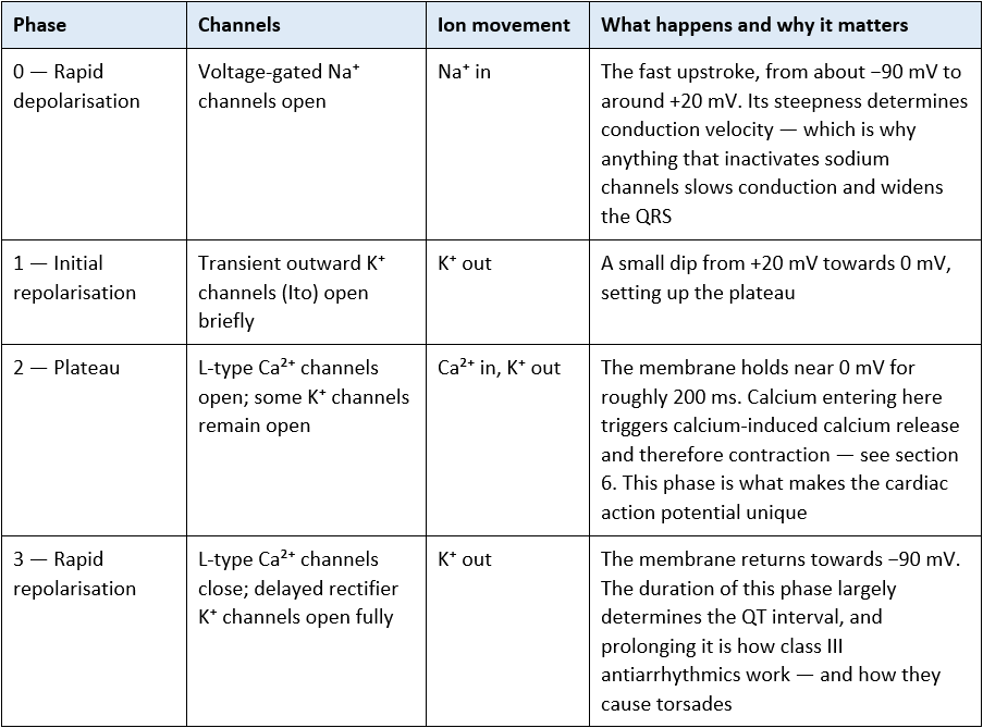
Key features
The long duration of the action potential prevents tetany — the muscle cannot be re-stimulated before it has relaxed and refilled.
Calcium influx during the plateau is essential for excitation–contraction coupling.
The effective refractory period lasts nearly the whole action potential, preventing premature re-excitation.
4. The pacemaker (slow response) action potential
Sinoatrial and atrioventricular nodal cells behave differently. Their job is not to conduct fast or contract — it is to fire spontaneously and, in the case of the AV node, to delay. Their action potential reflects that.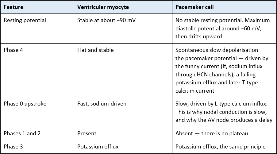
The rate at which a pacemaker cell fires depends on three things: the slope of phase 4, the threshold potential, and the maximum diastolic potential. Anything that changes one of those changes the heart rate.
Why this section matters clinically
Sympathetic stimulation (beta-1, via cAMP) steepens the slope of phase 4 — threshold is reached sooner, so the rate rises. Vagal stimulation (M2) hyperpolarises the cell and flattens the slope — the rate falls. This is the mechanism behind the bradycardia you see during suctioning and intubation.
Because the nodal upstroke is calcium-dependent rather than sodium-dependent, calcium channel blockers such as verapamil and diltiazem slow the sinus rate and AV conduction — while sodium channel blockers have little effect there.
Ivabradine blocks the funny current specifically, slowing the rate without any negative inotropic effect.
Adenosine hyper-polarises nodal cells and transiently blocks AV conduction — which is precisely why it is used.
See Cardiac Conduction System and Common Cardiac Drugs.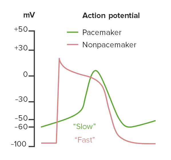
side-by-side comparison of the ventricular and pacemaker action potentials, with phase 4 slope highlighted on the pacemaker trace.
5. Refractory periods and why they protect the heart
Absolute refractory period — no stimulus of any strength can trigger another action potential, because the sodium channels are inactivated and have not yet reset. This covers phase 0 through most of phase 3.
Relative refractory period — a stronger than normal stimulus can trigger an action potential, but it will be slow-rising, low-amplitude and conducted poorly. This is late phase 3, and it is the vulnerable period.
The refractory period is long, covering nearly the entire action potential. That is what prevents the heart from being re-stimulated before it has relaxed and refilled, and therefore what prevents tetany. A skeletal muscle can be driven into sustained contraction; a heart cannot, and must not.
R on T — the vulnerable period in practice
The relative refractory period corresponds roughly to the T wave on the ECG. A stimulus landing here — an ectopic beat, a pacing spike, or an unsynchronised shock — can trigger ventricular tachycardia or ventricular fibrillation.
This is why cardioversion is synchronised.
The defibrillator waits for the R wave so the energy is not delivered on the T wave. If you have ever pressed shock and had nothing happen, the commonest reason is that synchronisation is on and the machine cannot find an R wave to trigger on.
It is also why defibrillation in ventricular fibrillation is deliberately unsynchronised — there is no organised R wave to synchronise to, and waiting for one would mean never delivering the shock.
And it is why a pacemaker that fails to sense is dangerous: a spike delivered during the T wave is an R-on-T event. See Principles of Cardiac Pacing.
6. Excitation–contraction coupling: from calcium to squeeze
Step 2 content — rationale and application
An action potential is only useful if it produces a contraction. The link between the two is calcium.
During phase 2, calcium enters through L-type channels in the T-tubules.
That relatively small amount of calcium triggers the release of a much larger store from the sarcoplasmic reticulum, through ryanodine receptors — calcium-induced calcium release.
Calcium binds troponin C, moving tropomyosin off the actin binding sites and allowing cross-bridge cycling. The cell shortens.
Relaxation requires calcium to be removed: most is pumped back into the sarcoplasmic reticulum by SERCA, and the remainder is extruded by the sodium–calcium exchanger, which moves three sodium ions in for every calcium ion out.
The amount of calcium available to troponin during each beat is what determines contractility. Every inotrope works by changing that number.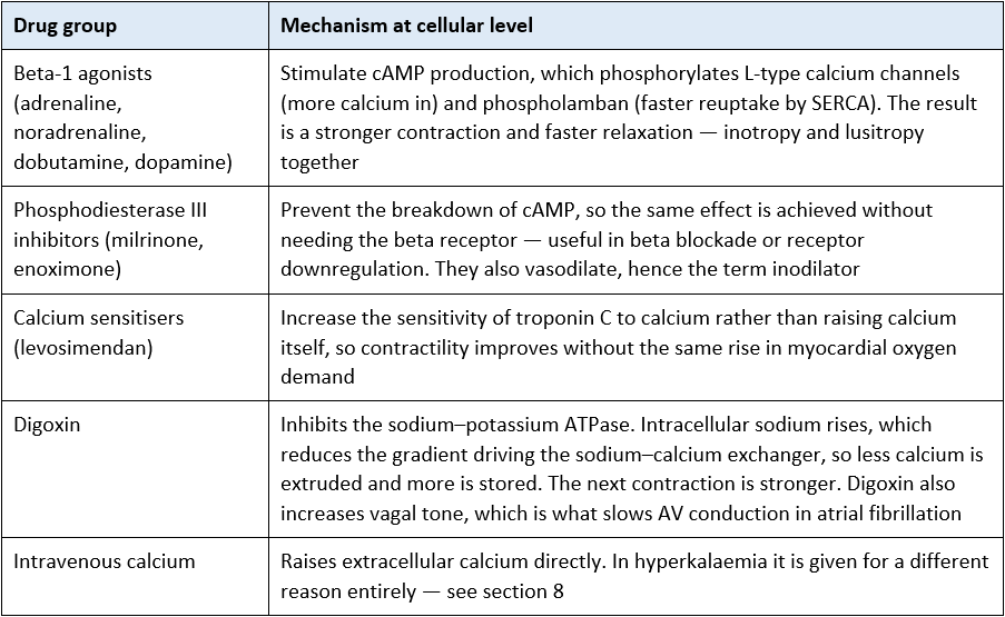
Digoxin and potassium — a high-yield nursing point
Potassium and digoxin compete for the same binding site on the sodium–potassium ATPase.
When potassium is low, more digoxin binds — so a patient can become clinically toxic at a digoxin level that is reported as being within range.
Hypomagnesaemia and hypercalcaemia also increase toxicity. Renal impairment raises the level itself, because digoxin is renally cleared.
In practice: check the potassium and magnesium alongside any digoxin level, and treat a normal level in a hypokalaemic patient with suspicion rather than reassurance.
7. Linking the action potential to the ECG
The ECG is the summed electrical activity of millions of these action potentials, recorded from the body surface. Each part of the complex corresponds to a phase.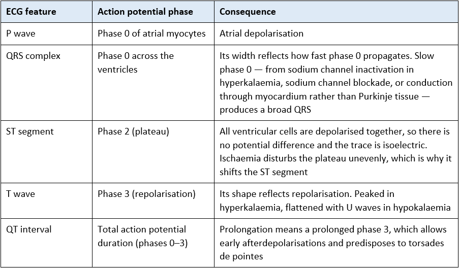
The reasoning shortcut: QRS width is a phase 0 problem — think sodium, conduction, potassium. QT length is a phase 3 problem — think potassium, magnesium, calcium and drugs. Once you can place an ECG abnormality on the action potential, the cause list writes itself.
See 12 lead ECG Essentials and Cardiac Conduction System.
8. Electrolytes and the action potential
8.1 Potassium
Hyperkalaemia. Raised extracellular potassium makes the resting membrane potential less negative, so the cell sits partially depolarised. Sodium channels progressively inactivate at this less negative potential, so phase 0 becomes slower and smaller — conduction slows. At the same time repolarisation is accelerated.
Peaked, tented T waves — accelerated phase 3.
Flattening then loss of P waves, and PR prolongation — atrial tissue and the AV node failing first.
Progressive QRS widening — sodium channel inactivation slowing phase 0.
Merging of the widened QRS with the T wave into a sine wave pattern, then ventricular fibrillation or asystole.
A widening QRS in a patient at risk of hyperkalaemia is a pre-arrest sign. Escalate immediately and follow your local hyperkalaemia protocol.
Why the treatments do what they do — ICU Step 2
Intravenous calcium (chloride or gluconate) removes no potassium at all. It raises the threshold potential away from the partially depolarised resting potential, restoring the gap between the two and stabilising the myocardium. It works within minutes and lasts around 30 to 60 minutes — it buys time, it does not treat the problem.
Insulin with dextrose drives potassium into cells by stimulating the sodium–potassium ATPase. It shifts potassium, it does not remove it — so monitor the glucose and expect the potassium to rebound.
Nebulised salbutamol shifts potassium intracellularly by the same beta-2 mediated stimulation of the ATPase.
Only dialysis, ion exchange binders and renal excretion actually remove potassium from the body.
This distinction — stabilise, shift, remove — is exactly the rationale a Step 2 assessor will ask you to give. See Acute Kidney Injury and Indications for Renal Replacement Therapy.
Hypokalaemia. Low extracellular potassium hyperpolarises the cell and prolongs repolarisation. On the ECG: flattened T waves, U waves, ST depression and an apparently prolonged QT. The prolonged phase 3 permits early afterdepolarisations, which is the mechanism of torsades de pointes. Hypokalaemia is also a potent contributor to ventricular ectopy and ventricular tachycardia in the critically ill.
8.2 Magnesium
Magnesium stabilises the membrane and shortens the QT. Hypomagnesaemia prolongs the QT and facilitates early after-depolarisations — and magnesium is the treatment for torsades de pointes.
Magnesium is a cofactor for the sodium–potassium ATPase. This is why refractory hypokalaemia will not correct however much potassium you give until the magnesium has been replaced — the pump that moves potassium into cells cannot work without it. Check and replace both together.
8.3 Calcium
Hypocalcaemia prolongs phase 2 and therefore the QT interval; hypercalcaemia shortens it.
Watch for hypocalcaemia in massive transfusion (citrate binds calcium) and in citrate-anticoagulated renal replacement therapy — both are common critical care situations with a direct effect on this trace.
8.4 Temperature and pH
Hypothermia slows all channel kinetics: bradycardia, prolonged PR, QRS and QT, and J (Osborn) waves.
Acidosis impairs channel function and contractility, and drives potassium out of cells, raising the serum level.
9. Drugs and the action potential
Step 2 content — rationale and application
The Vaughan Williams classification is simply a map of which phase each antiarrhythmic acts on.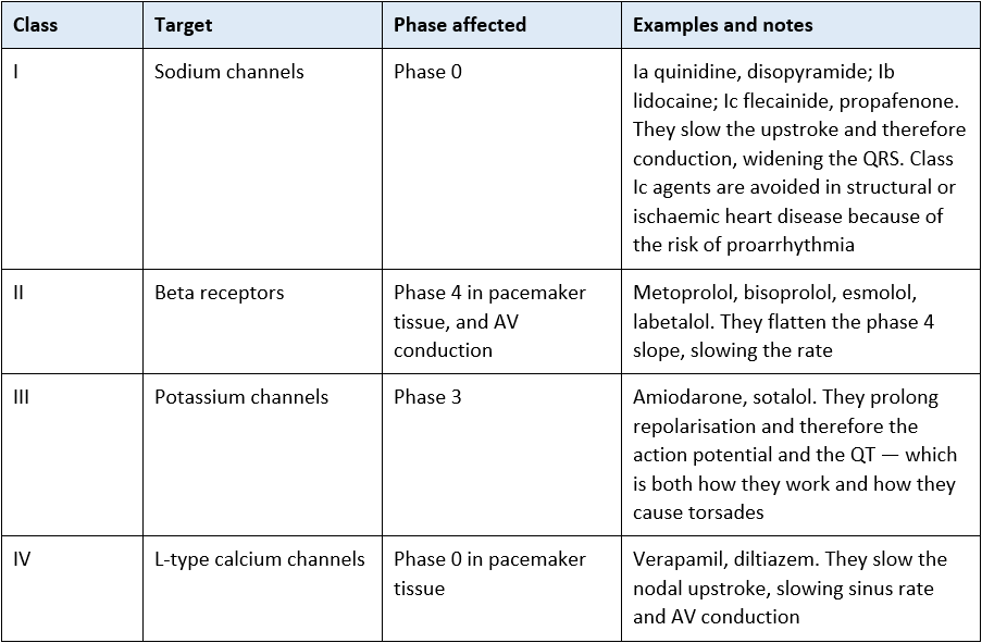
*Amiodarone is formally class III but has properties of all four classes, which is why it is so widely used and why it has so many effects and interactions.
**Adenosine, digoxin, magnesium and ivabradine sit outside the classification but are all explained by this physiology — see sections 4 and 6.
QT prolongation is cumulative
Many drugs used routinely in critical care prolong phase 3: amiodarone, sotalol, haloperidol and other antipsychotics, ondansetron, macrolides, quinolones and some antifungals.
The risk multiplies when several are prescribed together, and is amplified by hypokalaemia and hypomagnesaemia — which are also common in the same patients.
Before a new QT-prolonging drug is added, the QTc and the potassium and magnesium are all worth having to hand. Follow local QT monitoring guidance.
Two further groups worth knowing
Local anaesthetics are sodium channel blockers. Systemic toxicity therefore presents with conduction disturbance, broad complex arrhythmia and arrest, alongside neurological features. This is a recognised emergency with a specific treatment; know where your unit keeps the lipid emulsion and the AAGBI guideline.
Beta blocker and calcium channel blocker overdose produce profound bradycardia and hypotension that may be refractory to atropine. Specific treatments exist. Escalate early and involve the poisons service through TOXBASE.
10. Ischaemia, hypoxia and acidosis
Step 2 content — rationale and application
The sodium–potassium ATPase runs on ATP. When oxygen delivery fails, ATP falls, and the consequences cascade:
The pump slows. Sodium accumulates inside the cell, potassium leaks out into the extracellular space, and the resting potential becomes less negative.
Partially depolarised cells have slow, small phase 0 upstrokes and conduct poorly.
ATP-sensitive potassium channels open, shortening the action potential in the ischaemic zone but not in neighbouring healthy tissue.
The result is a patchwork of tissue with different action potential durations and different refractory periods side by side. That heterogeneity is the substrate for re-entry — and re-entry is the mechanism of most ventricular tachycardia and fibrillation.
Rising intracellular sodium also reverses the sodium–calcium exchanger, so calcium accumulates inside the cell, contributing to both arrhythmia and to contractile dysfunction.
This is why the first hour after coronary occlusion carries the highest risk of ventricular fibrillation, why reperfusion itself can be arrhythmogenic, and why correcting hypoxia, acidosis and electrolytes is antiarrhythmic treatment in its own right rather than supportive care around the edges of it.
11. Clinical implications for the acute care nurse
Every shift
Treat potassium and magnesium as cardiac observations, not as biochemistry. Know the current values on your patient and when they were last checked.
Replace magnesium alongside potassium, and expect potassium replacement to fail if you do not.
Look at the QRS width and the T wave shape when you review the rhythm, not just the rate.
Review the drug chart for accumulating QT-prolonging agents, particularly when a new antiemetic, antipsychotic or antibiotic is added.
When titrating
Understand what you are titrating towards and why: a beta agonist infusion is increasing calcium availability per beat, and doing so at the cost of increased myocardial oxygen demand.
Monitor for the predictable consequences — tachyarrhythmia, ectopy and ischaemia — and report them rather than simply increasing the rate.
Follow local policy and prescribed limits; titration outside those limits is not within scope, whatever the physiology suggests.
Escalate immediately for:
A widening QRS in a patient at risk of hyperkalaemia — a pre-arrest sign requiring the local protocol now, not after the next blood gas.
New QT prolongation, particularly with hypokalaemia, hypomagnesaemia or a newly added drug.
Polymorphic ventricular tachycardia — consider torsades, and know that magnesium is the treatment.
Suspected digoxin toxicity, including in a patient whose level is reported as within range but whose potassium is low.
Conduction disturbance or arrhythmia after local anaesthetic administration — think systemic toxicity.
12. Self-test
Check your understanding. The section to check your answer against is shown in brackets.
ICU Step 1
– Name the five phases of the ventricular action potential and the main ion movement in each. (Section 3)
– Which ion determines the resting membrane potential, and what maintains the gradient? (Section 2)
– Give three ways the pacemaker action potential differs from the ventricular one. (Section 4)
– What is the refractory period, and why does the heart need a long one? (Section 5)
– Which phase of the action potential does the QRS represent, and which does the T wave represent? (Section 7)
– Describe the sequence of ECG changes in worsening hyperkalaemia. At which point would you escalate? (Section 8.1)
– Why is cardioversion synchronised while defibrillation in VF is not? (Section 5)
Step 2
– Explain, using the action potential, why hyperkalaemia widens the QRS. (Sections 2, 3 and 8.1)
– Your patient with a potassium of 6.9 is given intravenous calcium. A colleague asks why, since the potassium has not changed. Answer them. (Section 8.1)
– Distinguish the mechanisms of calcium, insulin with dextrose, and nebulised salbutamol in hyperkalaemia using the terms stabilise, shift and remove. (Section 8.1)
– Your patient’s potassium will not correct despite repeated replacement. What else would you check and why? (Section 8.2)
– Explain how digoxin increases contractility, and why hypokalaemia makes a normal digoxin level unreliable. (Section 6)
– Map the four Vaughan Williams classes onto the phases of the action potential, with one example of each. (Section 9)
– Explain at cellular level why the first hour after coronary occlusion carries the highest risk of ventricular fibrillation. (Section 10)
– Why do class III antiarrhythmics both treat and cause arrhythmia? (Sections 3, 7 and 9)
Abbreviations
AAGBI Association of Anaesthetists of Great Britain and Ireland
ATP Adenosine triphosphate
ATPase Adenosine triphosphatase (the sodium–potassium pump)
AV Atrioventricular
cAMP Cyclic adenosine monophosphate
CC3N Critical Care Networks – National Nurse Leads
ECG Electrocardiogram
HCN Hyperpolarisation-activated cyclic nucleotide-gated (channel)
IK1 Inward rectifier potassium current
Ito Transient outward potassium current
QTc Corrected QT interval
SA Sinoatrial
SERCA Sarcoplasmic/endoplasmic reticulum calcium ATPase
VF / VT Ventricular fibrillation / ventricular tachycardia
Glossary
Depolarisation — the membrane potential becoming less negative, towards and beyond zero.
Repolarisation — the membrane potential returning towards its resting negative value.
Threshold potential — the membrane potential at which an action potential becomes self-sustaining.
Early after-depolarisation — an abnormal upward deflection during phase 3, favoured by a prolonged action potential, and the trigger for torsades de pointes.
Re-entry — a self-perpetuating circuit of electrical activity, arising where neighbouring tissue has differing conduction velocities and refractory periods.
Inotropy / lusitropy — the strength of contraction / the rate of relaxation.
Proarrhythmia — the capacity of an antiarrhythmic drug to cause the arrhythmia it is meant to prevent.
Tetany (in muscle) — a sustained contraction produced by rapidly repeated stimulation, which the long cardiac refractory period prevents.
Recommended pages
Next in this section
Cardiac Conduction System — how these action potentials travel through the heart
Cardiac v Skeletal Muscle — why cardiac muscle cannot be tetanised
The Cardiac Cycle — the mechanical consequence
Starling’s Law — the other determinant of contractility
Applying this clinically
12 lead ECG Essentials; Heart Rhythms – Overview; Management of Cardiac Arrhythmias
Common Cardiac Drugs — inotropes, vasopressors and antiarrhythmics
Acute Kidney Injury; Basic Considerations in Renal Failure; Indications for Renal Replacement Therapy — hyperkalaemia
Principles of Cardiac Pacing — sensing, capture and R on T
Cardiogenic Shock; Pathophysiology of Heart Failure
Refeeding Syndrome — electrolyte shifts and arrhythmia risk
Further reading and local governance
Guidelines
Resuscitation Council UK — adult advanced life support, including the peri-arrest algorithms and the treatment of hyperkalaemia in cardiac arrest.
UK Kidney Association — clinical practice guideline on the treatment of acute hyperkalaemia in adults.
AAGBI / Association of Anaesthetists — management of severe local anaesthetic toxicity.
TOXBASE — for beta blocker, calcium channel blocker and digoxin overdose.
NICE NG196 — Atrial fibrillation: diagnosis and management.
CC3N National Competency Framework for Registered Nurses in Adult Critical Care — Step 1 and Step 2
Local governance
Your local hyperkalaemia protocol and electrolyte replacement guidelines.
Your local QT monitoring guidance and high-risk drug combination list.
Your local inotrope and vasopressor prescribing and titration policy, and the limits within which you may titrate.
The location of your unit’s lipid emulsion and the local local-anaesthetic toxicity pathway.
Textbooks
Levick, An Introduction to Cardiovascular Physiology
Adam, Osborne and Welch, Critical Care Nursing: Science and Practice (Oxford)
Bersten and Handy, Oh’s Intensive Care Manual
Last reviewed: July 2026. Next review due: July 2027. AskAlth, London UK.
AskAlth is an independent educational resource created by NHS critical care nurses. It is not affiliated with CC3N, NICE, or the NHS, though our content is mapped to and referenced against their published frameworks and guidance.