

Coronary Artery Anatomy
How to use this resource, and learning outcomes:
1. Why coronary anatomy matters at the bedside
2. Origin of the coronary arteries
3. Left coronary artery (LCA)
4. Right coronary artery (RCA)
5. Coronary dominance
6. Blood supply to the conduction system
7. Linking anatomy to the 12-lead ECG
8. Clinical implications for the acute care nurse
Abbreviations
9. Test your knowledge
Further reading and local governance
How to use this resource
Work through sections 1 to 6 to build the anatomy, then use sections 7 and 8 to link that anatomy to what you see at the bedside: the ECG, the blood pressure, the rhythm and the patient in front of you.
Learning outcomes
By the end of this session you will be able to:
Describe the origin and course of the right and left coronary arteries and their major branches.
State which region of the myocardium each major artery supplies.
Explain coronary dominance and why it matters clinically.
Identify the blood supply to the sinoatrial (SA) and atrioventricular (AV) nodes.
Relate ECG territory changes to the likely culprit artery.
Explain why coronary perfusion is diastolic, and the implications for haemodynamic management.
1. Why coronary anatomy matters at the bedside
The coronary circulation delivers oxygen and nutrients to the heart muscle (myocardium). It arises from the ascending aorta, immediately above the aortic valve, as the right and left coronary arteries.
The myocardium extracts almost all of the oxygen delivered to it at rest. Unlike other tissues, the heart cannot meaningfully increase oxygen extraction when demand rises — it can only increase flow. Anything that limits flow (a stenosis, a thrombus, a low diastolic pressure) rapidly becomes ischaemia.
Coronary flow happens mainly in diastole. During systole the contracting myocardium compresses the vessels running through it, particularly on the left side. The left ventricle is therefore perfused almost entirely between beats.
Coronary perfusion pressure (CPP) is approximated as:
CPP ≈ aortic diastolic pressure − left ventricular end-diastolic pressure (LVEDP)
Practical consequences you will see:
Tachycardia shortens diastole disproportionately, reducing ventricular filling and coronary perfusion time. A rate of 150 is not only a rhythm problem; it is an ischaemia problem.
A low diastolic pressure reduces coronary perfusion — relevant in vasoplegic shock, in aortic regurgitation, and whenever you are titrating vasodilators.
A high LVEDP (fluid overload, poor left ventricular compliance) opposes coronary filling.
This is the basis of intra-aortic balloon pump (IABP) counterpulsation: diastolic augmentation raises coronary perfusion pressure while systolic unloading reduces myocardial oxygen demand.
2. Origin of the coronary arteries
Both main arteries arise from sinuses — small outpouchings in the aortic root — just above the aortic valve cusps.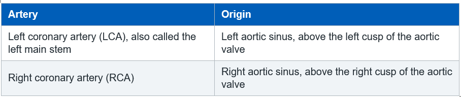
3. Left coronary artery (LCA)
Course: the left main stem is short. It passes between the pulmonary trunk and the left atrial appendage before dividing into its two main branches. In some people a third branch, the ramus intermedius, arises between them.
Because it supplies the majority of the left ventricle, occlusion of the left main stem is sometimes described as a “widow-maker” lesion. These patients are extremely unstable and are a resuscitation and rapid-transfer priority for cardiac intervention or surgery.
3.1 Left anterior descending artery (LAD)
Also called the anterior interventricular artery.
Course: runs in the anterior interventricular groove towards the apex of the heart.
Supplies:
Anterior wall of the left ventricle
Anterior two-thirds of the interventricular septum
Apex of the heart
Much of the bundle branch conducting tissue
Major branches:
Diagonal branches → anterolateral left ventricular wall
Septal perforators → anterior septum
3.2 Left circumflex artery (LCx)
Course: runs in the left atrioventricular (AV) groove, curving round towards the posterior aspect of the heart.
Supplies:
Lateral and posterior walls of the left ventricle
Left atrium
The SA node in approximately 40% of people
Major branches:
Obtuse marginal (OM) arteries → lateral left ventricular wall
In left-dominant circulation the LCx also gives rise to the posterior descending artery (see section 5).
4. Right coronary artery (RCA)
Course: runs in the right AV groove towards the crux cordis — the point on the posterior surface of the heart where the AV groove and the interventricular groove meet.
Supplies: the right atrium, the right ventricle and, in right-dominant hearts, the inferior wall of the left ventricle and the posterior septum.
Major branches: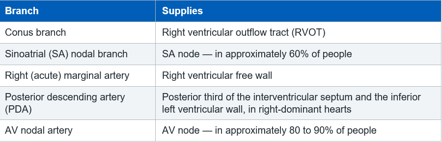
The RCA also supplies the posteromedial papillary muscle which, unlike its anterolateral counterpart, has only a single blood supply. This is why acute mitral regurgitation from papillary muscle rupture is classically a complication of inferior myocardial infarction.
5. Coronary dominance
Dominance describes which artery gives rise to the posterior descending artery (PDA). It does not describe which artery is larger or more important.
Percentages vary between anatomical sources. Use them as approximations rather than fixed figures.
Why this matters
In the majority (right-dominant) pattern, the RCA supplies both the AV node and the inferior wall. This is why inferior myocardial infarction so often presents with bradycardia and AV block.
In a left-dominant patient, an LCx occlusion can produce a far larger infarct than you might otherwise expect.
6. Blood supply to the conduction system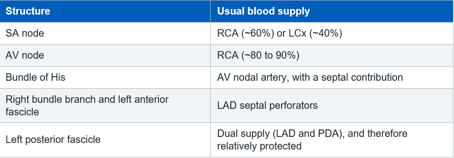
Bedside translation: conduction problems are territory-specific.
Inferior / RCA territory → sinus bradycardia, first-degree and Mobitz type I AV block. Usually transient, often responds to atropine and frequently resolves with reperfusion.
Anterior / LAD territory → new bundle branch block, Mobitz type II, complete heart block. This indicates a large septal infarct, carries a worse prognosis and often requires pacing.
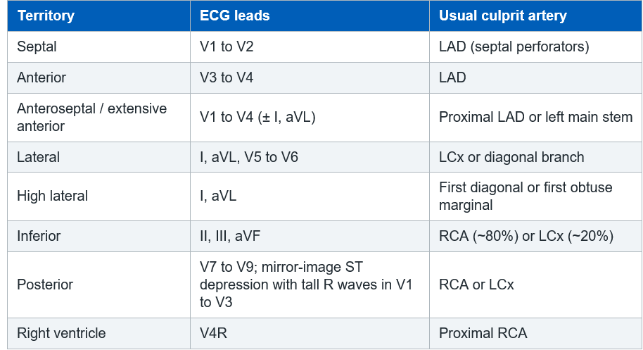
7. Linking anatomy to the 12-lead ECG
Practice point
ST elevation in the inferior leads should prompt a right-sided ECG (V4R) to look for right ventricular involvement.
Reciprocal ST depression in V1 to V3 alongside inferior changes should prompt posterior leads (V7 to V9).
8. Clinical implications for the acute care nurse
Right ventricular infarction (proximal RCA)
The right ventricle is thin-walled and preload-dependent. When it infarcts, cardiac output depends on adequate filling.
Classic picture: hypotension, raised jugular venous pressure, clear lung fields.
Nitrates and opiates can cause profound hypotension. Use with extreme caution and only after senior discussion.
Diuretics are generally contraindicated in the acute phase.
Management is usually cautious fluid loading with close reassessment, plus inotropic support if filling alone is insufficient.
Have atropine and external pacing immediately available.
Large anterior infarction (proximal LAD)
Anticipate cardiogenic shock, pulmonary oedema and malignant arrhythmias.
Mechanical complications to watch for: ventricular septal rupture, free wall rupture and left ventricular thrombus.
A new murmur, sudden deterioration or unexplained shock warrants urgent echocardiography.
Patients after PCI or CABG
Know which vessel was treated before you take handover. Your monitoring priorities differ between an inferior RCA lesion (rhythm and preload) and a proximal LAD lesion (pump failure).
Recurrent chest pain, ST re-elevation or a new arrhythmia after PCI should be escalated immediately — consider stent thrombosis.
Check and document the access site, distal pulses and limb perfusion.
Everyday monitoring priorities
Continuous ECG monitoring using an appropriate lead, with alarm limits set for the individual patient.
Balance oxygen supply and demand: treat pain, anxiety, fever, anaemia and tachyarrhythmia, all of which increase myocardial oxygen consumption.
Repeat the 12-lead ECG with any change in symptoms and compare it against the baseline.
9. Self-test
Complete these before your competency discussion. The section to check your answer against is shown in brackets.
– Which artery supplies the anterior two-thirds of the interventricular septum? (Section 3.1)
– Your patient has ST elevation in II, III and aVF and is bradycardic at 38 beats per minute. Which artery is most likely involved, and why is the bradycardia anatomically predictable? (Sections 4 and 6)
– What does “right dominant” mean, and how common is it? (Section 5)
– Why should you be cautious with GTN in a patient with a suspected right ventricular infarct? (Section 8)
– Your patient’s heart rate rises from 80 to 160 beats per minute. Give two reasons why this worsens myocardial ischaemia. (Section 1)
– Which additional leads would you record if you suspected a posterior infarct? (Section 7)
Abbreviations
AV Atrioventricular
CABG Coronary artery bypass graft
CPP Coronary perfusion pressure
GTN Glyceryl trinitrate
IABP Intra-aortic balloon pump
LAD Left anterior descending artery
LCA Left coronary artery
LCx Left circumflex artery
LV / LVEDP Left ventricle / left ventricular end-diastolic pressure
OM Obtuse marginal
PCI Percutaneous coronary intervention
PDA Posterior descending artery
RCA Right coronary artery
RVOT Right ventricular outflow tract
SA Sinoatrial
Related Pages:
Before this page
Structure and Function of the Heart — chambers, walls and layers
Cardiac Surface Anatomy — the grooves and the crux cordis these arteries run in
The Cardiac Cycle — why coronary filling happens in diastole
Alongside this page
Coronary Artery Structure and Histology — the vessel wall itself
Coronary Veins — how blood leaves the myocardium
Cardiac Conduction System — the tissue fed by the SA and AV nodal branches
Apply it at the bedside
12 lead ECG territories — matching the leads to the culprit artery
Coronary Artery Disease — what happens when these vessels narrow
Acute Coronary Syndrome — recognition and immediate management
Myocardial Infarction — territory, complications and treatment
Cardiogenic Shock — when pump failure follows a large infarct
Principles of Cardiac Pacing — for the heart block that follows RCA and LAD occlusion
Working through your ICU competencies? Follow the Step 1 ICU Pathway.
Further reading and local governance
CC3N National Competency Framework for Registered Nurses in Adult Critical Care, Step 1 — cardiovascular section.
Your local acute coronary syndrome, chest pain and cardiac arrest guidelines.
Resuscitation Council UK peri-arrest arrhythmia algorithms (bradycardia and tachycardia).
NICE guidance on acute coronary syndromes.
A current anatomy and physiology text for critical care nursing, for the underpinning science.
