

Acquired Heart Valve Disease — Overview
How to use this resource, and learning outcomes:
1. Why acquired valve disease matters in critical care
2. Definitions and mechanisms
3. Causes of acquired valve disease
4. The eight lesions at a glance
5. Acute versus chronic: the distinction that changes everything
6. Infective endocarditis
7. Assessing severity and the decision to intervene
8. Haemodynamic management principles in critical care
9. Monitoring caveats in valve disease
10. Interventions and post-procedure care
11. Prosthetic valves and anticoagulation
12. General management principles
13. Clinical implications for the acute care nurse
Abbreviations
14. Self-test
Glossary
Recommended pages
Further reading and local governance
What this page covers — and what it does not
This page covers the ABNORMAL valve: what goes wrong, why, how each lesion presents, how severity is graded, how these patients are managed in critical care, and what happens after surgical or transcatheter intervention.
For the NORMAL valve — valve anatomy, chordae and papillary muscles, the fibrous skeleton, why valves are passive, the aortic root, auscultation landmarks and normal heart sounds — see Heart Valves.
This page assumes that anatomy. If the terms coaptation, annulus, chordae tendineae or dicrotic notch are unfamiliar, start there.
How to use this resource
Section 4 is the reference table — use it to look up a lesion quickly. Sections 5 to 13 are the parts you need when the patient is in front of you: how acute lesions differ from chronic ones, how to recognise endocarditis, what the haemodynamic plan is trying to achieve, when your monitoring is misleading you, and what to watch for after intervention.
ICU Step Competencies:
If you are working through Step 1, sections 2 to 6, 12 and 13 are your priority.
If you are working through Step 2 or an academic critical care programme, sections 5, 7, 8, 9 and 10 are where the rationale sits — you will be asked to justify the targets, not just recite them.
Learning outcomes
Step 1 — by the end of this session you will be able to:
– Define stenosis and regurgitation and explain the haemodynamic consequence of each.
– Name the common causes of acquired valve disease in the UK.
– Describe the characteristic murmur, clinical features and typical treatment of the four left-sided lesions.
– Recognise the presentation of acute severe mitral or aortic regurgitation and escalate appropriately.
– List the features that should raise suspicion of infective endocarditis, and state what must be taken before antibiotics are started.
– State the current UK position on antibiotic prophylaxis for endocarditis.
– Describe the nursing priorities for a patient with a prosthetic valve.
Step 2 — in addition, you will be able to:
– Give a rationale for the haemodynamic targets used in each lesion, including heart rate, rhythm, preload and afterload.
– Explain why acute severe regurgitation behaves so differently from the chronic form, using ventricular and atrial compliance.
– Explain why intra-aortic balloon counterpulsation is contraindicated in aortic regurgitation.
– Identify when waveform interpretation and cardiac output measurement are unreliable because of a valve lesion.
– Describe the parameters used to grade severity and the principles determining when intervention is offered.
– Anticipate and explain the specific complications of surgical and transcatheter valve intervention, including conduction block.
1. Why acquired valve disease matters in critical care
Acquired heart valve disease refers to damage or dysfunction of the heart valves that develops after birth. It may cause stenosis (narrowing) or regurgitation (leakage) of one or more valves, affecting normal blood flow, collectively referred to as lesions.
Such patients reach critical care by several routes: decompensating from a chronic lesion; presenting acutely with a valve that has just failed; after cardiac surgery or a transcatheter procedure; with infective endocarditis; or with an unrelated critical illness in which a known valve lesion changes everything about their management.
The lesion changes the rules. A fluid bolus, a rate control drug or a vasopressor that helps one patient can arrest another. Knowing which lesion your patient has, and which direction the blood is going, is the difference between anticipating deterioration and being surprised by it.
Acute valve failure presents as shock. Papillary muscle rupture, prosthetic valve thrombosis and endocarditic leaflet destruction all arrive as sudden deterioration with pulmonary oedema and, sometimes, a new murmur. Recognising the pattern is a nursing skill and it changes the outcome.
2. Definitions and mechanisms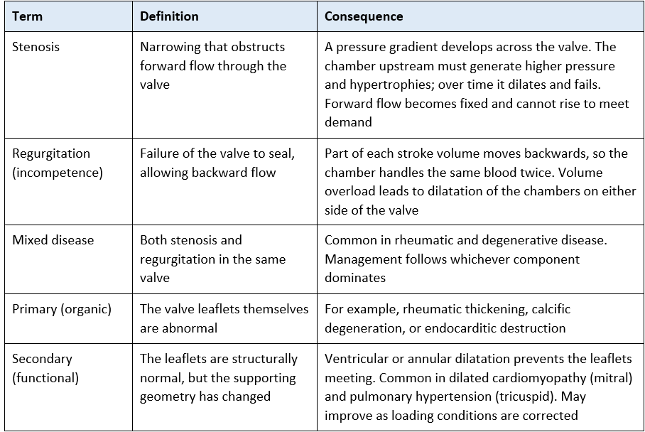
See Heart Valves for the normal valve anatomy and the fibrous skeleton — it explains why annular dilatation causes functional regurgitation with structurally normal leaflets.
3. Causes of acquired valve disease
Degenerative and calcific disease — the commonest cause in the UK and in ageing populations generally. Progressive fibrosis and calcification, particularly of the aortic valve and mitral annulus.
Rheumatic heart disease — an autoimmune consequence of group A streptococcal infection, developing years after the initial illness. Now uncommon as a new UK diagnosis but still the leading global cause, and frequently seen in older patients and in those who grew up outside the UK. It is by far the commonest cause of mitral stenosis.
Infective endocarditis — destroys leaflets and can perforate them, producing acute regurgitation. See section 6.
Ischaemic disease — papillary muscle dysfunction or rupture after myocardial infarction, causing acute mitral regurgitation. The posteromedial papillary muscle is vulnerable because it usually has only a single blood supply — see Heart Valves.
Myxomatous degeneration and prolapse — a common cause of chronic mitral regurgitation; chordal rupture can convert it acutely.
Chamber and annular dilatation — the mechanism of most functional mitral and tricuspid regurgitation seen in critical care.
Aortic root disease — root dilatation, connective tissue disorders and aortic dissection, all producing aortic regurgitation. Acute dissection involving the root is a surgical emergency.
Other — carcinoid syndrome (characteristically right-sided), radiotherapy, some drugs, and previous valve surgery with subsequent prosthetic dysfunction.
A note on bicuspid aortic valve
A bicuspid aortic valve is congenital rather than acquired — present in roughly 1 to 2% of the population.
It is included here because the resulting stenosis is acquired: the abnormal valve degenerates and calcifies earlier than a normal one, making it a leading cause of aortic stenosis in adults under 65.
It is also associated with aortopathy, so these patients may have aortic root or ascending aortic dilatation as well.
4. The eight lesions at a glance
Use this as a reference table. The four left-sided lesions are the ones you will meet most often in adult critical care.
Left-sided heart lesions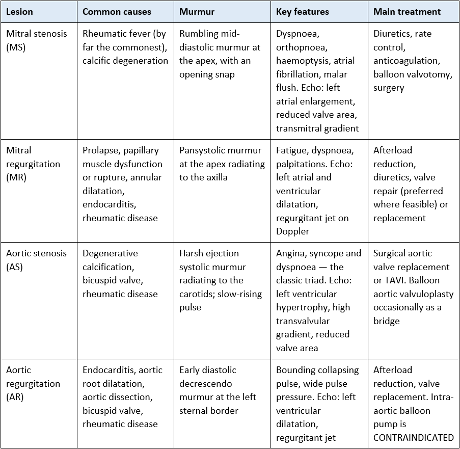
Right-sided heart lesions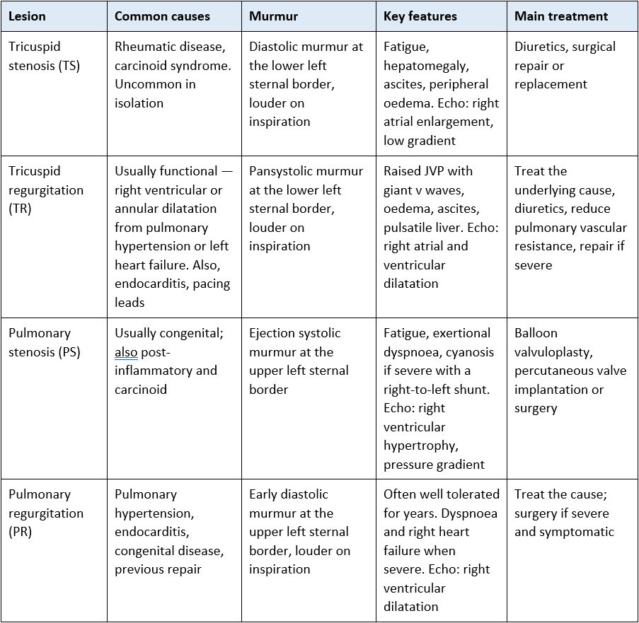
Right-sided murmurs and inspiration
Right-sided murmurs typically get louder on inspiration; left-sided murmurs do not.
The reason is simple physiology: inspiration lowers intrathoracic pressure, increasing venous return to the right heart, so more blood flows across the right-sided valves and the murmur intensifies.
This is a useful bedside discriminator and a favourite question at competency discussion. For where to listen and how, see Heart Valves and Heart Sounds (auscultation).
5. Acute versus chronic: the distinction that changes everything
Step 2 content — rationale and application
The same named lesion behaves as two entirely different diseases depending on how fast it developed. This is the single most important framing for critical care, because the acute forms are the ones that arrive on your unit in extremis.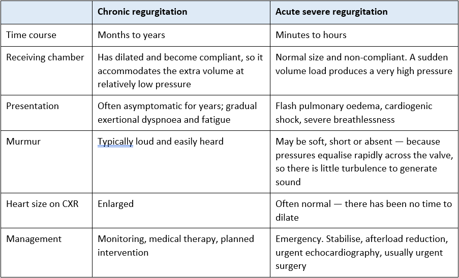
The trap — do not be reassured by a quiet chest or a normal chest film
In acute severe mitral regurgitation, the murmur can be soft or absent despite catastrophic regurgitation, and the heart can look normal size on the radiograph.
A patient with sudden severe breathlessness and shock two to seven days after an inferior myocardial infarction has papillary muscle rupture until proven otherwise — regardless of how quiet their chest sounds.
Acute severe aortic regurgitation from endocarditis or aortic dissection behaves the same way and is equally surgical.
Escalate for urgent echocardiography. Do not wait for the murmur.
6. Infective endocarditis
Endocarditis is both a cause of acquired valve disease and one of the commonest reasons a valve patient reaches critical care. Because valve leaflets are largely avascular, they have limited immune surveillance and poor antibiotic penetration — which is why infection can establish there and why antibiotics alone often cannot clear it.
6.1 When to suspect it
– Fever or unexplained sepsis in a patient with any prosthetic valve, known valve disease, a cardiac device or a history of endocarditis.
– A new or changed regurgitant murmur.
– Fever in a person who injects drugs — typically right-sided, affecting the tricuspid valve, with septic pulmonary emboli.
– Embolic phenomena: stroke, splinter haemorrhages, splenic or renal infarction, mycotic aneurysm.
– Prolonged unexplained fever with a recent line, dental work, or invasive procedure.
6.2 Diagnosis
Blood cultures are the priority — the recommended number of sets from separate sites, taken before antibiotics wherever the patient’s condition allows. Once antibiotics have started, cultures may never be positive and the diagnosis becomes far harder. Follow local policy.
Echocardiography: transthoracic first, with transoesophageal imaging where suspicion is high or the transthoracic study is inadequate — particularly for prosthetic valves and for root abscess.
Diagnosis uses established international criteria combining microbiological, imaging and clinical findings.
6.3 Complications to watch for
Acute regurgitation and heart failure — the commonest indication for surgery and the commonest cause of death.
New conduction block — a red flag. The AV node and bundle of His lie immediately adjacent to the aortic valve, so new PR prolongation or heart block suggests a root abscess extending into the conduction tissue. Escalate urgently. See Cardiac Conduction System.
Embolic events — stroke is the most feared. Perform and document regular neurological observations.
Persistent fever or positive cultures on treatment — suggests uncontrolled infection and may indicate surgery.
Prophylaxis — the UK position
– NICE CG64 does not recommend routine antibiotic prophylaxis against infective endocarditis before dental or other procedures for people at risk.
– Instead, patients should be supported with good oral health, told which symptoms to report, and advised of the risks of non-sterile procedures such as body piercing and tattooing.
– Some individual patients are managed differently on specialist advice. Follow local policy, and note that practice in other countries differs.
– In practice, meticulous asepsis and intravascular device care prevent far more endocarditis than any prophylactic antibiotic.
6.4 Nursing priorities
Cultures before antibiotics, documented accurately with times and sites.
Prolonged intravenous antibiotics — protect the line, monitor for complications, and support the patient through what is often a six-week course.
Daily neurological observations and a low threshold for escalating any new deficit.
Continuous ECG monitoring with attention to the PR interval and any new block.
Temperature trend, inflammatory markers and repeat cultures as prescribed.
Screening for the source — dental review, line review, skin inspection.
7. Assessing severity and the decision to intervene
Step 2 content — rationale and application
Severity is assessed by echocardiography, using several parameters together rather than any single number. You do not need to perform the assessment, but you should be able to read the report and understand what it is describing.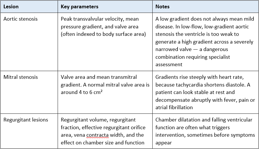
What drives the decision to intervene
Symptoms. In aortic stenosis in particular, the onset of angina, syncope or breathlessness marks a clear prognostic watershed — outcomes without intervention deteriorate sharply from that point.
Objective severity on imaging.
Ventricular size and function — intervening before irreversible ventricular damage is a central principle.
Whether the patient is undergoing cardiac surgery for another reason.
Procedural risk, frailty, comorbidity and — importantly — the patient’s own priorities.
Decisions are made by a multidisciplinary heart team. The 2025 ESC/EACTS guidelines moved intervention earlier in the disease course and expanded the role of transcatheter therapies, including at younger ages than previously. If you are supporting a patient through this decision, your role is to make sure their questions and priorities reach the team.
8. Haemodynamic management principles in critical care
Step 2 content — rationale and application
These are physiological principles to help you understand the plan and anticipate deterioration — not a treatment protocol. Always follow local policy and the direction of the treating team.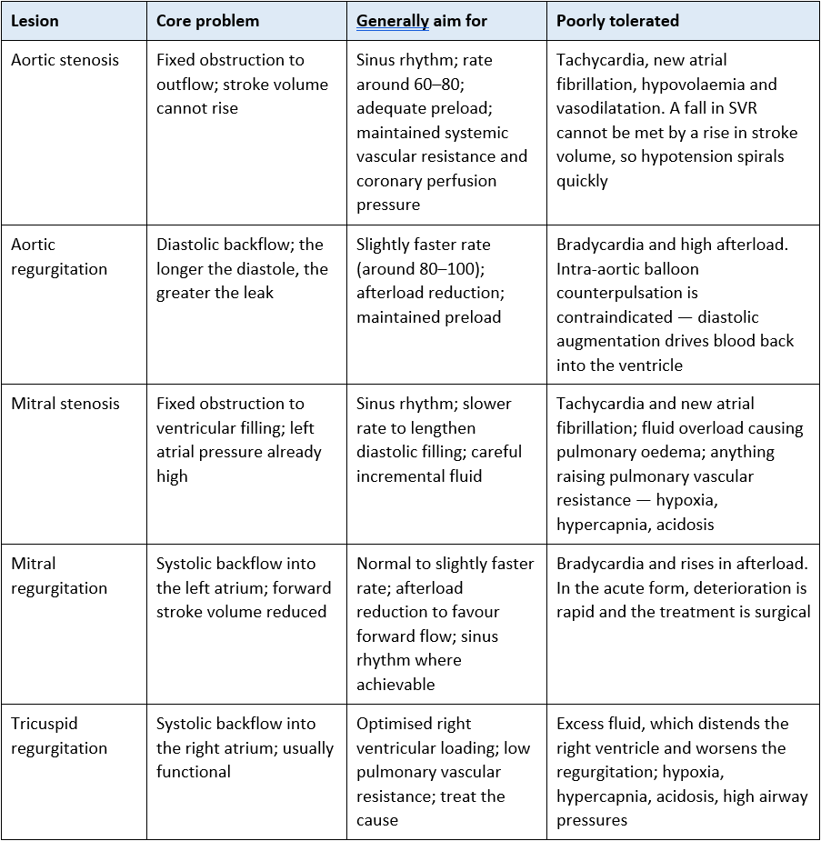
The pattern worth memorising:
Stenotic lesions generally want time — a slower rate and a longer diastole — and adequate preload. Regurgitant lesions generally want the load taken off and do not want to be slow. Everything else follows from asking which way the blood is going and what is stopping it.
Ventilation and fluid — two recurring themes
Positive pressure ventilation reduces preload and left ventricular afterload. It is badly tolerated in preload-dependent, rate-sensitive lesions — induction and intubation in severe aortic or mitral stenosis is a recognised cause of profound hypotension and arrest. Anticipate it and have vasopressor ready.
It is helpful in acute pulmonary oedema from acute mitral regurgitation, where non-invasive ventilation reduces both preload and afterload.
Give fluid in small aliquots and reassess against a response rather than a target number. In stenotic lesions and hypertrophied ventricles the pressure–volume relationship is steep: the ventricle needs filling but congests with comparatively little volume.
For the underlying physiology see Heart Valves (valve loading and ventilation) and Starling’s Law (fluid responsiveness, which holds the fullest treatment of bolus volumes and reassessment).
9. Monitoring caveats in valve disease
Step 2 content — rationale and application
Valve lesions distort the waveforms you rely on, and in some cases make cardiac output measurement unreliable. Knowing when not to trust a number is as much a Step 2 skill as knowing how to read one.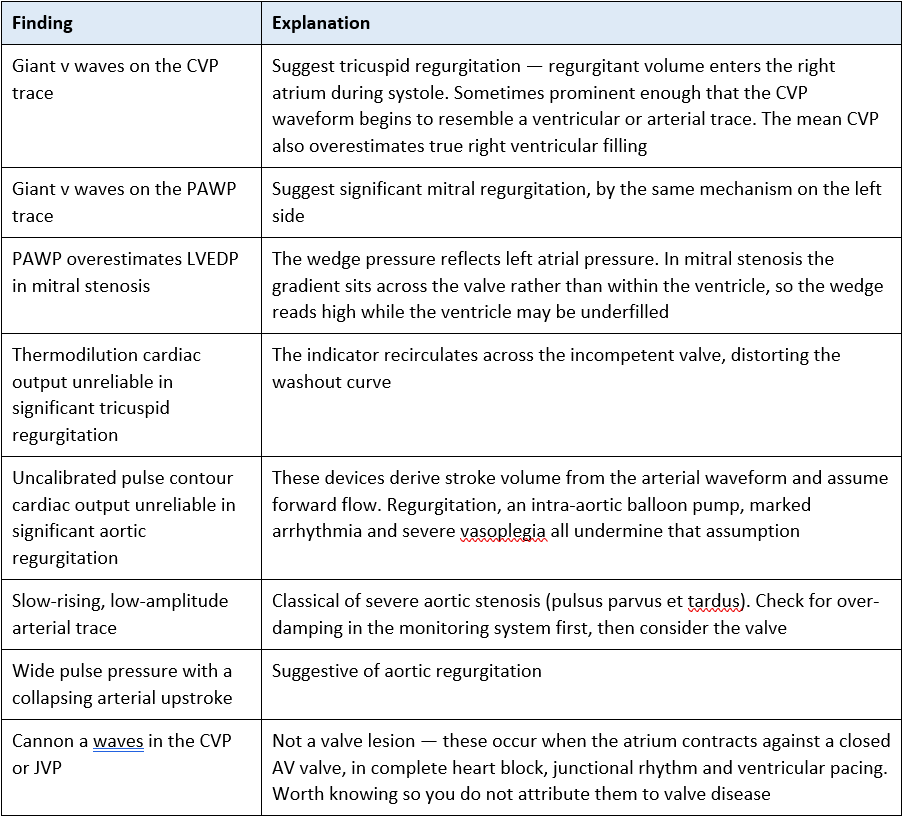
For the normal arterial and CVP waveform components and what each represents, see Heart Valves section 9. For monitoring technique and troubleshooting, see Invasive Monitoring – Waveforms and Cardiac Output Monitoring.
10. Interventions and post-procedure care
10.1 The options
Surgical repair — preferred over replacement for the mitral valve wherever feasible, because it preserves the patient’s own valve apparatus and avoids prosthetic anticoagulation.
Surgical replacement — with a mechanical or bioprosthetic valve. See section 11.
TAVI — transcatheter aortic valve implantation, now offered at progressively younger ages under the 2025 ESC/EACTS guidance.
Transcatheter mitral and tricuspid procedures — including edge-to-edge repair, for selected patients at high surgical risk.
Balloon valvotomy or valvuloplasty — for suitable mitral and pulmonary stenosis, and occasionally as a bridge in aortic stenosis in a patient too unstable for definitive treatment.
10.2 After cardiac surgery
Bleeding and tamponade — a sudden fall in mediastinal drain output alongside deteriorating haemodynamics is a red flag; the drains may have clotted. Hypotension, raised filling pressures, tachycardia and a rising lactate should prompt immediate escalation and preparation for emergency resternotomy in line with local policy.
Conduction disturbance — particularly after aortic valve surgery, because of the proximity of the conduction tissue to the aortic valve. Epicardial pacing wires are usually in place; confirm from the operation note which are atrial and which are ventricular.
Post-operative atrial fibrillation — very common, and poorly tolerated in a stiff or recently operated ventricle. Correct potassium and magnesium and escalate promptly.
Vasoplegia after cardiopulmonary bypass — low systemic vascular resistance with an adequate cardiac output, often requiring vasopressor support.
Routine recovery priorities — analgesia, early extubation where appropriate, glycaemic control, mobilisation and restarting anticoagulation to the surgical plan.
10.3 After TAVI
– New conduction block is a recognised complication and complete heart block requiring permanent pacing is well described — these patients stay on cardiac monitoring afterwards.
– Vascular access complications: bleeding, haematoma, pseudoaneurysm and limb ischaemia. Check the site and distal perfusion.
– Stroke, paravalvular leak and acute kidney injury from contrast.
See Epicardial Pacing following Cardiac Surgery and Principles of Cardiac Pacing.
11. Prosthetic valves and anticoagulation
11.1 Mechanical valves
Durable, but require lifelong anticoagulation with a vitamin K antagonist, usually warfarin. Direct oral anticoagulants are contraindicated in mechanical valves.
The target INR depends on the valve type, its position and patient risk factors. Take it from the cardiology or surgical plan rather than assuming a standard range, and check the plan for periprocedural bridging.
Expect a crisp metallic click. A newly muffled or absent click warrants urgent review — consider valve thrombosis or pannus, particularly if anticoagulation has been interrupted.
Chronic low-grade haemolysis is expected. Suspect significant haemolysis with falling haemoglobin, raised LDH, raised unconjugated bilirubin, raised reticulocytes and low haptoglobin — report it, as it may indicate a paravalvular leak.
11.2 Bioprosthetic valves
Most do not require long-term anticoagulation, though a short initial period of anticoagulant or antiplatelet therapy is common. Follow the surgical plan.
Structural degeneration occurs over roughly 10 to 20 years, and sooner in younger patients — which is part of the trade-off discussed when the valve type is chosen.
Interrupted anticoagulation is a recurring critical care problem
– Valve patients frequently have anticoagulation held for bleeding, procedures or invasive lines.
– Every interruption carries thrombotic risk, and for a mechanical valve that risk includes valve thrombosis and systemic embolism.
– Make sure the plan for stopping and restarting is explicit, documented and handed over, and escalate if it is unclear or has drifted.
12. General management principles
– Control symptoms — for example diuretics for congestion, with careful attention to not over-diuresing a preload-dependent patient.
– Manage arrhythmias, especially atrial fibrillation, which is both common and poorly tolerated in stenotic and stiff-ventricle lesions.
– Anticoagulate where indicated — for atrial fibrillation, for mechanical valves and after certain procedures, according to the individual plan.
– Prevent complications through meticulous asepsis, intravascular device care and support with oral health — rather than through routine antibiotic prophylaxis, which is not recommended in the UK. See section 6.
– Correct the reversible drivers of decompensation: anaemia, infection, hypoxia, pain, fever, thyroid disease and electrolyte disturbance.
– Ensure timely surgical or interventional correction before irreversible ventricular damage develops — the central principle running through all valve guidance.
13. Clinical implications for the acute care nurse
At handover
– Establish which valve lesion or prosthesis the patient has, its severity, and whether it is native, repaired or replaced.
– Establish the agreed haemodynamic targets — rate, rhythm, mean arterial pressure, filling — and the reasoning behind them.
– Establish the anticoagulation plan, INR target where relevant, and when the last dose and level were.
– Note the baseline murmur, or the documented absence of one, and the character of any prosthetic click, if confident to do so.
Through the shift
– Protect rhythm. New atrial fibrillation in aortic stenosis, mitral stenosis or a hypertrophied ventricle is a haemodynamic emergency and needs to be managed carefully.
– Correct the reversible drivers of arrhythmia and of raised pulmonary vascular resistance: pain, anxiety, fever, hypoxia, hypercapnia, acidosis, hypovolaemia, anaemia, potassium and magnesium.
– Give fluid in small aliquots and reassess against response rather than a single pressure number.
– Know which of your monitoring numbers this patient’s lesion makes unreliable — see section 9.
– Take blood cultures before antibiotics in any febrile valve or prosthesis patient.
Escalate immediately for:
Sudden deterioration with a new murmur — consider acute severe regurgitation from papillary muscle or chordal rupture, or endocarditic destruction.
Flash pulmonary oedema with cardiogenic shock, particularly two to seven days after an inferior myocardial infarction.
A muffled, changed or absent prosthetic click, especially with subtherapeutic or interrupted anticoagulation.
Unexplained fever in a valve or prosthesis patient — cultures before antibiotics.
New conduction block, after valve surgery or TAVI, or in suspected endocarditis where it may indicate root abscess.
A sudden fall in mediastinal drain output with deteriorating haemodynamics after cardiac surgery.
14. Self-test
Check your knowledge. The section to check your answer against is shown in brackets.
Step 1
– Define stenosis and regurgitation, and state the haemodynamic consequence of each. (Section 2)
– Name the commonest cause of mitral stenosis worldwide, and the commonest cause of aortic stenosis in the UK. (Section 3)
– Describe the murmur of aortic stenosis, where it radiates, and the classic triad of symptoms. (Section 4)
– Why do right-sided murmurs get louder on inspiration? (Section 4)
– What must be taken before antibiotics are started in a febrile patient with a prosthetic valve, and why does it matter? (Section 6)
– Is routine antibiotic prophylaxis before dental work recommended in the UK for patients at risk of endocarditis? (Section 6)
– Your patient with a mechanical valve has a newly muffled click. What are you worried about? (Section 11)
Step 2
– Explain why acute severe mitral regurgitation presents so differently from the chronic form, and why the murmur may be soft. (Section 5)
– Why is an intra-aortic balloon pump contraindicated in aortic regurgitation? (Sections 4 and 8)
– Give a rationale for aiming for a slower heart rate in mitral stenosis and a faster one in aortic regurgitation. (Section 8)
– Your patient with severe aortic stenosis is about to be induced and ventilated. Predict what will happen and explain why. (Section 8)
– The CVP trace on your patient with tricuspid regurgitation looks almost arterial and the thermodilution cardiac output readings are scattered. Explain both findings. (Section 9)
– Why is the wedge pressure a poor guide to left ventricular filling in mitral stenosis? (Section 9)
– A patient being treated for endocarditis develops first-degree heart block. Why does this worry you? (Section 6.3)
– Explain how a patient with severe aortic stenosis can have a low transvalvular gradient. (Section 7)
– Your post-aortic-valve-replacement patient becomes hypotensive and tachycardic, and the mediastinal drain that was draining 60 mL/hr has stopped. What is your working diagnosis and what are your immediate actions? (Section 10.2)
Abbreviations
AF – Atrial fibrillation
AR / AS – Aortic regurgitation / aortic stenosis
AVR – Aortic valve replacement
CC3N – Critical Care Networks – National Nurse Leads
CXR – Chest radiograph
ESC / EACTS – European Society of Cardiology / European Association for Cardio-Thoracic Surgery
IABP – Intra-aortic balloon pump
INR – International normalised ratio
JVP – Jugular venous pressure
LDH – Lactate dehydrogenase
LVEDP – Left ventricular end-diastolic pressure
MR / MS – Mitral regurgitation / mitral stenosis
PAWP – Pulmonary artery wedge pressure
PR / PS – Pulmonary regurgitation / pulmonary stenosis
PVR / SVR – Pulmonary / systemic vascular resistance
TAVI – Transcatheter aortic valve implantation
TR / TS – Tricuspid regurgitation / tricuspid stenosis
TOE / TTE – Transoesophageal / transthoracic echocardiography
Glossary
Coaptation — the meeting and sealing of valve leaflets when closed.
Opening snap — a sharp early diastolic sound produced by a stenotic but still mobile mitral valve.
Paravalvular leak — regurgitation around the outside of a prosthetic valve, between the sewing ring and the annulus.
Pannus — fibrous tissue ingrowth that can obstruct a prosthetic valve.
Pulsus parvus et tardus — the slow-rising, low-amplitude pulse of severe aortic stenosis.
Regurgitant fraction — the proportion of stroke volume flowing backwards rather than forwards.
Root abscess — an infected cavity in the aortic root, a complication of endocarditis which may involve the conduction tissue.
Vegetation — an infected mass of platelets, fibrin and organisms on a valve.
Vena contracta — the narrowest point of a regurgitant jet, used in grading severity.
Recommended pages
Underpinning anatomy and physiology
Heart Valves — the companion page. Normal valve anatomy, chordae and papillary muscles, the fibrous skeleton, pressure gradients, the aortic root, auscultation landmarks and normal waveform components
Heart Sounds (auscultation) — technique in detail
The Cardiac Cycle; Starling’s Law; Cardiac Conduction System
Applying this clinically
Cardiogenic Shock; Left Ventricular Failure; Pathophysiology of Heart Failure
Management of Cardiac Arrhythmias; Principles of Cardiac Pacing; Epicardial Pacing following Cardiac Surgery
Invasive Monitoring – Waveforms; Cardiac Output Monitoring; Arterial Line Transducer
Fluid Management – Colloid v Crystalloid; Common Cardiac Drugs
Cardiac Tamponade; Myocardial Infarction; Acute Coronary Syndrome
Hypertrophic (Obstructive) Cardiomyopathy; Dilated Cardiomyopathy; Cor Pulmonale
Further reading and local governance
Guidelines
2025 ESC/EACTS Guidelines for the Management of Valvular Heart Disease — the current standard, replacing the 2021 edition.
NICE NG208 — Heart valve disease presenting in adults: investigation and management.
NICE CG64 — Prophylaxis against infective endocarditis.
ESC 2023 Guidelines on the Management of Infective Endocarditis.
NICE NG196 — Atrial fibrillation: diagnosis and management.
British Society of Echocardiography — minimum datasets and valve assessment protocols.
CC3N National Competency Framework for Registered Nurses in Adult Critical Care — Step 1 and Step 2.
Local governance
Your local anticoagulation, warfarin bridging and INR reversal policies.
Your local blood culture policy — number of sets, sites and timing.
Your local cardiac surgery emergency resternotomy policy, if applicable to your unit.
Your local post-TAVI and post-cardiac-surgery monitoring standards.
Your local endocarditis pathway and outpatient parenteral antibiotic service arrangements.
Textbooks
Adam, Osborne and Welch, Critical Care Nursing: Science and Practice (Oxford)
Bersten and Handy, Oh’s Intensive Care Manual
Last reviewed: August 2026. Next review due: August 2027. AskAlth Nursing Team, London UK.
AskAlth is an educational resource for registered healthcare professionals in UK adult critical care. Not for patients or the public. Not a substitute for local policy, clinical guidance or the BNF. Content reflects guidance at the time of writing. AskAlth is independent and not affiliated with CC3N, NICE, the Resuscitation Council UK, the NMC or the NHS.
AskAlth is an independent educational resource created by NHS doctors and critical care nurses. It is not affiliated with CC3N, NICE, or the NHS, though our content is mapped to and referenced against their published frameworks and guidance.