

Difference Between Cardiac and Skeletal Muscle
How to use this resource, and learning outcomes:
1. Why this comparison matters at the bedside
2. The three muscle types at a glance
3. Structure: intercalated discs and the functional syncytium
4. Control: automaticity versus neural stimulation
5. Grading force: why the heart cannot recruit or tetanise
6. Calcium: the difference that explains the drugs
7. Metabolism and blood supply: why the heart cannot tolerate an oxygen debt
8. Regeneration and repair
9. What this means for drugs used in critical care
10. Skeletal muscle in critical illness
11. Biomarkers: troponin and creatine kinase
12. Clinical implications for the acute care nurse
Abbreviations
13. Self-test
Glossary
Recommended pages
Further reading and local governance
How to use this resource
Sections 2 to 8 build the comparison. Sections 9 to 12 apply it — to the drugs you give, the weakness you will see in every long-stay patient, and the blood results you chase.
ICU Step ompetencies
If you are working through Step 1, sections 2 to 8, 11 and 12 are your priority.
If you are working through Step 2 or an academic critical care programme, sections 5, 6, 9 and 10 are where the rationale sits — this is the page that explains why a neuromuscular blocking agent paralyses the diaphragm but not the heart, and why a critically ill patient loses skeletal muscle so fast.
This page underpins the rehabilitation, mobility and sedation competencies in both Steps.
Learning outcomes
Step 1 — by the end of this session you will be able to:
-Compare cardiac, skeletal and smooth muscle by location, control, structure, contraction and regeneration.
– Explain what intercalated discs are and why they make the heart a functional syncytium.
– Explain why cardiac muscle cannot be tetanised and why that is essential.
– State how the heart increases its force of contraction, given that it cannot recruit more fibres.
– Explain why the heart depends on continuous coronary blood flow rather than on increased oxygen extraction.
– State why a raised troponin does not always mean myocardial infarction.
Step 2 — in addition, you will be able to:
– Explain why calcium channel blockers reduce cardiac contractility but do not weaken skeletal muscle.
– Explain why neuromuscular blocking agents paralyse the diaphragm but leave the heart beating, and state the safety implication.
– Describe the mechanism by which suxamethonium can cause dangerous hyperkalaemia in specific patient groups.
– Describe ICU-acquired weakness and ventilator-induced diaphragmatic dysfunction, and give a rationale for early mobilisation and sedation minimisation.
– Distinguish rhabdomyolysis from myocardial injury using the pattern of creatine kinase and troponin.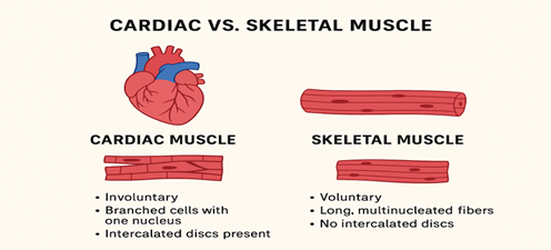 “>
“>
1. Why this comparison matters at the bedside
This is not a biology exercise. The differences between these tissues explain several things you deal with every shift.
Why the heart cannot be ‘worked harder’ the way a bicep can. Skeletal muscle increases force by recruiting more fibres and firing faster. The heart cannot do either. It has exactly two levers — fill it more, or make each contraction stronger — and those are the two things every haemodynamic intervention targets.
Why a paralysed patient still has a heartbeat. Neuromuscular blockers (‘paralysing agents’) act at a neuro junction that cardiac muscle does not have. The corollary is the important one: a fully paralysed patient can be fully awake (always therefore ensure patients who are paralysed have sufficient sedation).
Why long-stay patients become deconditioned while their hearts remains strong. Skeletal muscle is lost rapidly in critical illness — including the diaphragm, which is why it affects weaning — but it can regenerate. Cardiac muscle largely cannot, which is why an infarct leaves a scar and a scar leads to heart failure.
2. The three muscle types at a glance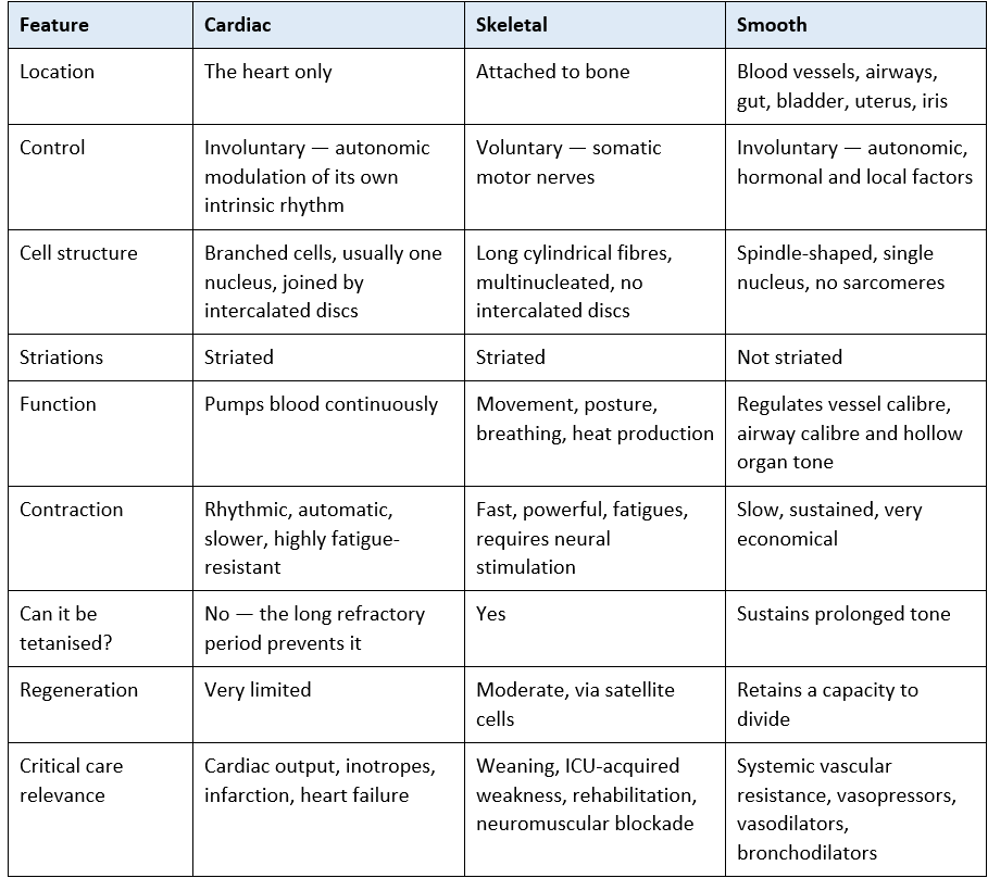 Features of Cardiac Skeletal Smooth muscle
“>
Smooth muscle is included because it matters.
Every vasopressor, vasodilator and bronchodilator you give acts on smooth muscle, and systemic vascular resistance — one of the two determinants of blood pressure — is simply the aggregate tone of the vascular smooth muscle in your patient’s arterioles.
3. Structure: intercalated discs and the functional syncytium
Intercalated discs are the specialised junctions where cardiac cells meet end to end. They contain two distinct types of connection:
(i) Gap junctions — low-resistance channels that allow ions, and therefore electrical current, to pass directly from one cell to the next.
(ii) Desmosomes and fascia adherens — mechanical anchors that hold the cells together and transmit the force of contraction from one cell to the next without the tissue tearing apart.
The functional syncytium
Because of the gap junctions, the myocardium behaves electrically as if it were a single cell. An impulse arriving anywhere spreads to the whole chamber.
The atria form one such syncytium and the ventricles another. The fibrous skeleton insulates them from each other, so the only normal route between them is the bundle of His — see the Cardiac Conduction System and Heart Valves pages.
The consequence is that the heart contracts as a unit, all or nothing. There is no such thing as contracting half a ventricle a bit harder. Skeletal muscle is the opposite. Its fibres are electrically independent, each supplied by its own motor neurone, and are recruited in graded fashion.
4. Control: automaticity versus neural stimulation
Skeletal muscle does nothing without a nerve. The motor neurone releases acetylcholine at the neuromuscular junction, acting on nicotinic receptors. Cut the nerve, block the junction, or lose the anterior horn cell, and the muscle is silent.
Cardiac muscle generates its own rhythm. Pacemaker cells depolarise spontaneously. The autonomic nervous system modulates the rate and force but does not initiate the beat. A denervated heart — a transplanted heart, for example — still beats, though it responds differently to stress and to drugs that work through the autonomic system.
The clinically vital consequence: cardiac muscle has no neuromuscular junction. Neuromuscular blocking agents therefore paralyse every skeletal muscle including the diaphragm, while the heart continues entirely unaffected. This is why a paralysed patient must always be adequately sedated — paralysis removes every outward sign of distress, including the ability to move, grimace or trigger the ventilator, while leaving awareness completely intact. See section 9.
5. Grading force: why the heart cannot recruit or tetanise
This section is the point of the whole comparison.
How skeletal muscle increases force
Recruitment — activating more motor units.
Rate coding and summation — firing faster, so contractions fuse before the muscle has relaxed.
At high enough frequency the contractions fuse completely into tetany, a sustained maximal contraction.
Why the heart can do neither
It is a functional syncytium, so every fibre is already recruited with every beat. There is nothing held in reserve to switch on.
Its refractory period lasts almost the entire action potential, so the muscle cannot be re-stimulated before it has relaxed and refilled. Summation, and therefore tetany, is impossible.
Why this is essential rather than a limitation
A tetanic heart would be a heart that never relaxes. A ventricle that does not relax does not fill, and a ventricle that does not fill ejects nothing.
Sustained contraction would abolish cardiac output within seconds — which, functionally, is what ventricular fibrillation is.
The long refractory period is therefore a protective mechanism, not an inefficiency. See Cardiac Action Potential.
So the heart has exactly two levers, and every haemodynamic intervention pulls one of them: preload — stretching the muscle more before it contracts (Starling’s law), and contractility — making each contraction stronger for the same stretch (inotropes). Rate is a third variable, but it changes output rather than the force of an individual beat, and beyond a point it reduces filling time and therefore reduces output. See Starling’s Law.
6. Calcium: the difference that explains the drugs
Step 2 content — rationale and application
Both muscle types contract when calcium binds troponin C. They differ in where that calcium comes from, and this single difference explains a surprising amount of pharmacology.
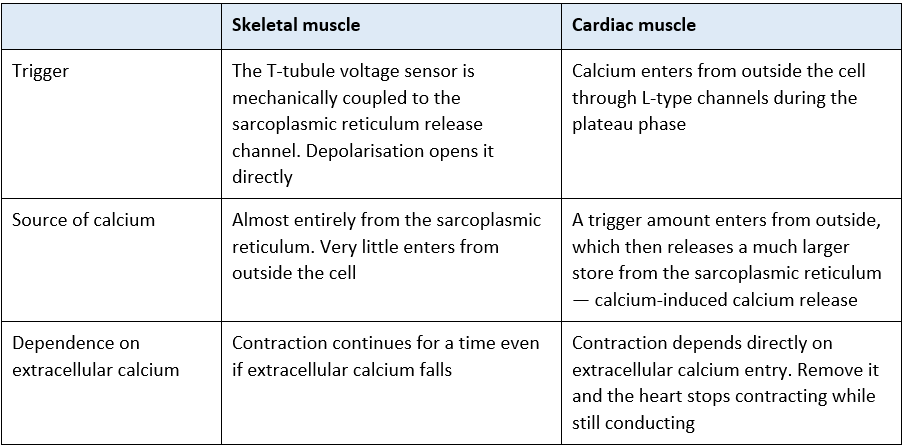 Calcium-skeletal-and-cardiac-muscle”>
Three consequences worth holding on to
Calcium channel blockers reduce cardiac contractility and slow nodal conduction, but do not cause skeletal muscle weakness — because skeletal muscle does not depend on that calcium influx.
Electrolyte disturbances hit the heart first. Potassium, calcium and magnesium abnormalities produce cardiac effects at concentrations that leave skeletal muscle largely functional. See Cardiac Action Potential.
Inotropes work by changing calcium handling. Beta agonists and phosphodiesterase inhibitors increase calcium availability; levosimendan increases the sensitivity of troponin C to the calcium already there. Starling’s law does something similar mechanically — stretch increases that same sensitivity.
7. Metabolism and blood supply: why the heart cannot tolerate an oxygen debt
Cardiac muscle is built for endurance. Roughly a third of its cell volume is mitochondria, it has a dense capillary network, and it is almost entirely aerobic — which is why it is described as fatigue-resistant.
But fatigue-resistant is not the same as tireless, and the distinction matters. Skeletal muscle can work anaerobically, build an oxygen debt, ache, and repay it afterwards. Cardiac muscle cannot. It has almost no anaerobic reserve, and when oxygen supply fails it stops working within minutes — first losing contractility, then becoming ischaemic, then infarcting.
Extraction is already maximal
At rest, the myocardium extracts roughly two-thirds to three-quarters of the oxygen delivered to it. Most other tissues extract around a quarter.
That means the usual compensation for increased demand — extracting more from the blood that is already arriving — is not available to the heart. It has almost nothing left to extract.
The only way the heart can meet increased demand is by increasing coronary blood flow.
This is the whole mechanism of angina and of demand ischaemia: whenever flow cannot rise to meet demand — because of a stenosis, tachycardia shortening diastole, hypotension reducing coronary perfusion pressure, anaemia, or hypoxia — the muscle becomes ischaemic.
It is also why tachycardia is doubly harmful: it raises demand and shortens the diastolic window in which the left coronary tree actually fills. See Coronary Artery Anatomy and Stable Angina.
8. Regeneration and repair
Skeletal muscle regenerates. Satellite cells sit alongside the fibres and can proliferate to repair damage and build new muscle. This is why rehabilitation after critical illness works, and why the wasting you see is, at least in part, recoverable.
Cardiac muscle largely does not. Turnover of cardiomyocytes in adult life is very low and falls further with age — nowhere near enough to replace the millions of cells lost in an infarct.
Damaged myocardium is therefore replaced by fibrous scar. Scar does not contract and does not conduct normally, which produces the two long-term consequences you will meet constantly: impaired contractility leading to ventricular remodelling and heart failure, and abnormal conduction providing the substrate for re-entrant arrhythmia.
The asymmetry is worth stating plainly: skeletal muscle wastes but can be rebuilt; cardiac muscle does not waste in the same way but cannot be rebuilt. It is the reason time matters so much in acute coronary syndrome — muscle saved in the first hours is muscle kept for life. See Myocardial Infarction and Pathophysiology of Heart Failure.
9. What this means for drugs used in critical care
Step 2 content — rationale and application
9.1 Neuromuscular blocking agents
These act at the nicotinic receptor of the skeletal neuromuscular junction. Cardiac muscle has no such junction and is unaffected. Smooth muscle is unaffected too — the gut and bladder keep working.
Safety essentials with neuromuscular blockade
Paralysis is not sedation and is not analgesia. A paralysed patient can be fully awake, in pain and terrified, with no way to signal it. Adequate sedation must be established before the blocker is given and maintained throughout.
The usual signs of inadequate sedation — movement, grimacing, ventilator dyssynchrony, coughing on suction — are all abolished. Sedation scoring tools are not valid in a paralysed patient. Unexplained tachycardia, hypertension, lacrimation or sweating may be the only clues, and they are unreliable.
Eye care matters: the blink reflex is lost and corneal abrasion is a real and preventable injury.
Airway and ventilator disconnection are immediately life-threatening, because the patient cannot breathe at all and cannot signal.
Peripheral nerve stimulator monitoring, where used locally, guides depth of blockade. Follow local policy.
9.2 Suxamethonium and extrajunctional receptors
Suxamethonium is a depolarising blocker: it opens the receptor and holds it open, which releases potassium from skeletal muscle. In a normal patient the resulting rise in serum potassium is small and unimportant.
In patients who have upregulated extrajunctional acetylcholine receptors, the release is massive and can cause cardiac arrest. Upregulation develops over roughly 24 to 72 hours and persists for months in: major burns, spinal cord injury, denervation and stroke, prolonged immobility, and prolonged critical illness — which includes a great many of the patients on your unit. Suxamethonium is contraindicated in these groups.
Suxamethonium is also a trigger for malignant hyperthermia, a skeletal muscle disorder of the ryanodine receptor, treated with dantrolene. The heart is not the primary tissue involved. Know where your unit’s dantrolene is kept.
9.3 Drugs acting on the other muscle types
Vasopressors and vasodilators act on vascular smooth muscle to change systemic vascular resistance and therefore blood pressure.
Bronchodilators act on airway smooth muscle.
Inotropes act on cardiac muscle calcium handling — see section 6.
Knowing which tissue a drug targets tells you what to monitor and what side effects to expect. See Common Cardiac Drugs and Common Medication used in Respiratory Care.
10. Skeletal muscle in critical illness
Step 2 content — rationale and application
This is where a critical care nurse actually applies skeletal muscle physiology, and it is the half of this topic most often left out.
10.1 ICU-acquired weakness
Critical illness polyneuropathy and critical illness myopathy frequently coexist, producing symmetrical limb weakness that becomes apparent as sedation is lightened. Recognised contributors include sepsis and systemic inflammation, multi-organ failure, immobility and bed rest, hyperglycaemia, and prolonged neuromuscular blockade; the role of corticosteroids remains debated.
Muscle loss begins early and is rapid, and is greatest in patients with multi-organ failure.
The consequences are prolonged ventilation, longer stay, and physical impairment that can persist for months or years after discharge.
10.2 The diaphragm
The diaphragm is skeletal muscle, and it wastes like any other skeletal muscle that is not used.
Under fully controlled mechanical ventilation the diaphragm does no work, and measurable thinning has been demonstrated within days. This is ventilator-induced diaphragmatic dysfunction, and it is one reason a patient who looks otherwise ready to wean fails a spontaneous breathing trial.
It is part of the rationale for minimising sedation, favouring modes that allow the patient to do some of the work, and avoiding unnecessary controlled ventilation.
It is also why weaning failure should prompt a question about muscle, not only about lungs and secretions. See Ventilator Weaning and Extubation – Overview.
10.3 What helps
Early mobilisation and rehabilitation, started as soon as it is safe, and progressed daily.
Minimising and interrupting sedation where clinically appropriate, so the patient can participate.
Adequate and appropriately timed nutrition, avoiding both underfeeding and overfeeding. See Nutrition in Critical Illness.
Glycaemic control within locally agreed targets.
Using neuromuscular blockade only where indicated, and reviewing the indication daily.
Setting rehabilitation goals with the patient and family, and documenting progress against them. See NICE CG83.
11. Biomarkers: troponin and creatine kinase
Because the two muscle types express different proteins, blood tests can distinguish which tissue has been damaged.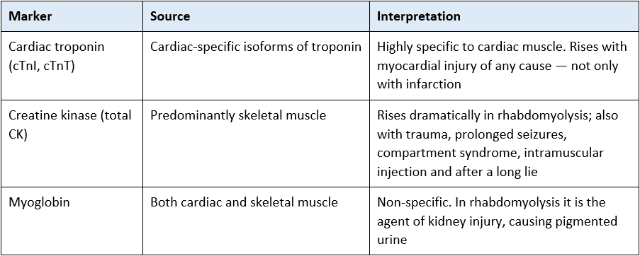
Two important points
A raised troponin does not mean myocardial infarction. It means myocardial injury. Sepsis, pulmonary embolism, arrhythmia, heart failure, kidney injury, cardiac contusion and critical illness itself all raise troponin. The diagnosis of infarction requires the clinical picture, ECG and the pattern of change — not a single number. Reporting the trend and the context is more useful than reporting the value.
In rhabdomyolysis, the CK is enormously raised while the troponin is normal or only mildly raised — a disproportion that tells you the damaged tissue is skeletal, not cardiac. The urgent issues are hyperkalaemia, acute kidney injury and fluid management. See Acute Kidney Injury.
12. Clinical implications for the acute care nurse
Cardiac muscle
– Remember the heart has only two levers. When asked whether to give fluid or start an inotrope, you are being asked which lever to pull — see Starling’s Law.
– Treat tachycardia in a patient with coronary disease as a demand problem as well as a rhythm problem: it raises oxygen requirement and shortens the diastolic filling window at the same time.
– Protect coronary perfusion pressure — diastolic pressure matters here, not just the mean.
– Correct anaemia, hypoxia and hypotension promptly in ischaemic patients; all three reduce oxygen delivery to a muscle that cannot compensate by extracting more.
Skeletal muscle
– Treat mobilisation as a clinical intervention with a dose, not as something done when there is time.
– Review sedation depth daily and advocate for the lightest safe level.
– Ask about muscle when a patient fails a weaning trial, not only about lungs and secretions.
– Position, splint and passively move limbs to prevent contracture in patients who cannot move themselves
Escalate for:
Any suggestion of awareness under neuromuscular blockade — and treat unexplained tachycardia, hypertension, sweating or lacrimation in a paralysed patient as inadequate sedation until proven otherwise.
A proposal to give suxamethonium to a patient with burns, spinal cord injury, denervation or prolonged immobility — raise it before the drug is drawn up.
A markedly raised CK with dark urine — think rhabdomyolysis, hyperkalaemia and kidney injury.
New symmetrical weakness as sedation lightens — flag it for medical and physiotherapy review rather than assuming it is simply deconditioning.
13. Self-test
Test your understanding. The section to check your answer against is shown in brackets.
Step 1
Give five differences between cardiac and skeletal muscle. (Section 2)
What are intercalated discs, and what are the two types of connection they contain? (Section 3)
What is meant by a functional syncytium, and why does it matter? (Section 3)
Skeletal muscle increases force by recruiting more fibres. How does the heart increase force, given that it cannot? (Section 5)
Why can cardiac muscle not be tetanised, and why would tetany be fatal? (Section 5)
Why must the heart increase coronary blood flow to meet increased demand, rather than extracting more oxygen? (Section 7)
Why does an infarct leave a scar while a torn hamstring heals? (Section 8)
Your patient’s troponin is raised but they have no chest pain and a normal ECG. Does this mean they have had a heart attack? (Section 11)
Step 2
Explain why verapamil reduces cardiac contractility but does not make a patient weak. (Section 6)
A colleague asks why the heart keeps beating in a fully paralysed patient. Answer them, and state the safety implication that follows. (Sections 4 and 9.1)
Which patients should not receive suxamethonium, and what is the mechanism of the danger? (Section 9.2)
Your patient has been fully ventilated and deeply sedated for eight days and has now failed two spontaneous breathing trials with clear lungs. Give a muscle-based explanation and three things that might have reduced the risk. (Section 10)
A patient found on the floor after a long lie has a CK of 40,000 and a troponin of 30. What is the likely diagnosis and what are your priorities? (Section 11)
How does Starling’s law relate to the calcium mechanism described in section 6? (Sections 5 and 6)
Why is tachycardia particularly dangerous in a patient with coronary artery disease? Give two mechanisms. (Sections 7 and 12)
Abbreviations
ACh – Acetylcholine
AKI – Acute kidney injury
CC3N – Critical Care Networks – National Nurse Leads
CK – Creatine kinase
cTnI / cTnT – Cardiac troponin I / cardiac troponin T
ICU-AW – Intensive care unit-acquired weakness
NMB – Neuromuscular blocking agent
NMJ – Neuromuscular junction
SR – Sarcoplasmic reticulum
SVR – Systemic vascular resistance
VIDD – Ventilator-induced diaphragmatic dysfunction
Glossary
Functional syncytium — a group of cells electrically coupled so that they behave as a single unit.
Gap junction — a channel allowing ions and current to pass directly between adjacent cells.
Motor unit — a single motor neurone and all the skeletal muscle fibres it supplies.
Recruitment — increasing force by activating more motor units; not available to cardiac muscle.
Satellite cell — a skeletal muscle stem cell capable of repairing and replacing damaged fibres.
Tetany (in muscle) — a sustained contraction produced by rapidly repeated stimulation.
Extrajunctional receptors — acetylcholine receptors that appear across the muscle membrane rather than only at the junction, following denervation, burns or prolonged immobility.
Remodelling — the change in ventricular size, shape and function that follows myocardial damage.
Recommended pages
Next in this section
Cardiac Action Potential — the refractory period and excitation–contraction coupling
Starling’s Law — the preload lever
Cardiac Conduction System — the syncytium in action
Structure and Function of the Heart; Heart Valves
Applying this clinically
Common Cardiac Drugs — inotropes, vasopressors and vasodilators
Coronary Artery Anatomy; Stable Angina; Myocardial Infarction; Pathophysiology of Heart Failure
Sedation and delirium assessment and management
Ventilator Weaning and Extubation – Overview; Basic Modes of Ventilation
Nutrition in Critical Illness; Patient Positioning
Acute Kidney Injury — rhabdomyolysis and hyperkalaemia
Further reading and local governance
Guidelines
NICE CG83 — Rehabilitation after critical illness in adults.
NICE QS158 — Rehabilitation after critical illness quality standard.
Faculty of Intensive Care Medicine / Intensive Care Society — Guidelines for the Provision of Intensive Care Services (GPICS), rehabilitation and sedation sections.
Association of Anaesthetists — guidance on neuromuscular blockade monitoring, and on malignant hyperthermia.
Fourth Universal Definition of Myocardial Infarction — for the distinction between myocardial injury and infarction.
CC3N National Competency Framework for Registered Nurses in Adult Critical Care
Local governance
Your local sedation, analgesia and neuromuscular blockade policy, including sedation scoring and peripheral nerve stimulator use.
Your local rapid sequence induction policy and its guidance on suxamethonium contraindications.
The location of your unit’s dantrolene and your local malignant hyperthermia pathway.
Your local early mobilisation and rehabilitation pathway, and rehabilitation goal documentation.
Your local rhabdomyolysis and troponin escalation pathways.
Textbooks
Tortora and Derrickson, Principles of Anatomy and Physiology — muscle tissue chapter
Klabunde, Cardiovascular Physiology Concepts (companion site: cvphysiology.com)
Adam, Osborne and Welch, Critical Care Nursing: Science and Practice (Oxford)
Bersten and Handy, Oh’s Intensive Care Manual
Last reviewed: August 2026. Next review due: August 2027. AskAlth Nursing Team, London UK.
AskAlth is an educational resource for registered healthcare professionals in UK adult critical care. Not for patients or the public. Not a substitute for local policy, clinical guidance or the BNF. Content reflects guidance at the time of writing. AskAlth is independent and not affiliated with CC3N, NICE, the Resuscitation Council UK, the NMC or the NHS.
AskAlth is an independent educational resource created by NHS critical care nurses. It is not affiliated with CC3N, NICE, or the NHS, though our content is mapped to and referenced against their published frameworks and guidance.